
Detección precoz de anemia de células falciformes por medio de un diagnóstico clínico y radiográfico oral. Reporte de caso / Early Detection of Sickle Cell Anemia by oral Diagnosis and oral Radiographs. Case Report


##plugins.themes.bootstrap3.article.details##
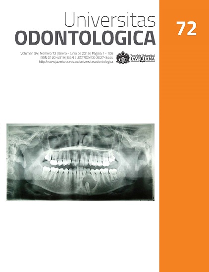
Antecedentes: La anemia de células falciformes (ACF) o drepanocitosis es una enfermedad mortal, diagnosticada regularmente en la adolescencia. No existen políticas de tamizaje temprano de ACF en Colombia. Objetivo: Reportar un caso de diagnóstico temprano de ACF por análisis clínico y radiográfico oral. Reporte del caso: A la consulta de la clínica odontológica de la Universidad Antonio Nariño (Popayán, Colombia) llega una paciente de etnia negroide de 9 años de edad, por sangrado en sus encías. En la valoración clínica, el índice de placa de O’Leary fue del 56,25 %. Se observó palidez palatal y sangrado al sondaje. Radiográficamente, en el maxilar inferior se observaron áreas multiloculares interradiculares en incisivos y primer molar izquierdo; además, había un patrón de escalera en el trabeculado óseo. Se ordenaron un hemograma y un extendido periférico que confirmaron hipocromía ligera y células falciformes positivas. Conclusiones: Aunque las manifestaciones clínicas y radiográficas orales no son patognomónicas de ACF, son una herramienta útil para realizar un diagnóstico temprano en odontología y ayudar a mejorar la calidad y esperanza de vida de los pacientes.
Background: Sickle cell anemia (SCA) or drepanocytosis is a fatal disease that is often diagnosed during adolescence. In Colombia, there are not early screening policies for SCA. Purpose: Report a case of SCA that was early diagnosed through clinical examination and radiographs. Case report: A 9-year-old African-Colombian girl attended the Antonio Nariño University dental clinic in Popayan, Colombia, with bleeding gums. The clinical examination showed 56.25 % dental plaque (O’Leary Index), palatal paleness, and bleeding after probing. The radiographic analysis evidenced multilocular zones between incisor roots and left first molar, with a staircase trabecular pattern in the bone. Findings from hemogram and peripheral blood analysis showed light hypochromia and positive sickle cells. Conclusion: Even though oral clinical and radiographic findings are not pathognomonic of SCA, they are a useful to early diagnose and improve people quality of life and life expectancy.
2. Musumadi L, Westerdale N, Appleby H. An overview of the effects of sickle cell disease in adolescents. Nurs Stand. 2012; 26(26): 35-40.
3. Silva JR, Malambo D, Silva DF, Fals E, Fals O, Rey J. Tamizaje de hemoglobinopatías en una muestra de la población infantil de Cartagena. Pediatr (Bogotá). 2003; 33(2).
4. Jaramillo M, Sáenz I, Pereira F. Tamizaje para anemia de células falciformes en recién nacidos del Hospital Universitario del Valle y del Hospital Mario Correa Renjifo. Actual Pediatr. 1997; 7(1): 3-13.
5. Domínguez Y, Zurita C, Calvopina D, Villacis J, Mora M. Prevalence of common hemoglobin variants in an Afro-descendent Ecuadorian population. BMC Res Notes. 2013; 6: 132.
6. Ballas SK, Gupta K, Adams-Graves P. Sickle cell pain: a critical reappraisal. Blood. 2012; 120(18): 3647-56.
7. Freitas LG, Isaac DL, Tannure WT, Lima EV, Abud MB, Tavares RS, Freitas CA, Avila MP. Retinal manifestations in patients with sickle cell disease referred to a University Eye Hospital. Arq Bras Oftalmol. 2011; 74(5): 335-7.
8. Maurer BT. A jaundiced view: more than meets the clinician’s eye. JAAPA. 2009; 22(10): 62.
9. Narang S, Fernandez ID, Chin N, Lerner N, Weinberg GA. Bacteremia in children with sickle hemoglobinopathies. J Pediatr Hematol Oncol. 2012; 34(1): 13-6.
10. MacMullen NJ, Dulski LA. Perinatal implications of sickle cell disease. MCN Am J Matern Child Nurs. 2011; 36(4): 232-8.
11. Benavides J, García H. Priapismo y anemia de células falciformes: una revisión de la literatura. Rev Urol Colomb. 2013; 22(2): 37-42.
12. Granados A, Hernández O, Guerra A, Uribe C. Moyamoya syndrome and neurological complications in a patient with sickle cell disease. Acta Neurol Colomb. 2012; 28(1): 49-54.
13. Marti-Carvajal AJ, Knight-Madden JM, Martinez-Zapata MJ. Interventions for treating leg ulcers in people with sickle cell disease. Cochrane Database Syst Rev. 2012; 11: Cd008394.
14. Muriel A. Drepanocitosis en embarazo. Rev Colomb Salud Libre. 2008; 3(2): 176-85.
15. Nwadiaro HC, Ugwu BT, Legbo JN. Chronic osteomyelitis in patients with sickle cell disease. East Afr Med J. 2000; 77(1): 23-6.
16. Sadat-Ali M. The status of acute osteomyelitis in sickle cell disease: A 15-year review. Int Surg. 1998; 83(1): 84-7.
17. Marti-Carvajal AJ, Agreda-Perez LH. Antibiotics for treating osteomyelitis in people with sickle cell disease. Cochrane Database Syst Rev. 2012; 12: Cd007175.
18. Stevenson H, Boardman C, Chu P, Field A. Mental nerve anesthesia: A complication of sickle cell crisis during childbirth. Dent Update. 2004; 31(8): 486-7.
19. Hamdoun E, Davis L, McCrary SJ, Eklund NP, Evans OB. Bilateral mental nerve neuropathy in an adolescent during sickle cell crises. J Child Neurol. 2012; 27(8): 1038-41.
20. Costa CP, Thomaz EB, Souza Sde F. Association between sickle cell anemia and pulp necrosis. J Endod. 2013; 39(2): 177-81.
21. Madani G, Papadopoulou AM, Holloway B, Robins A, Davis J, Murray D. The radiological manifestations of sickle cell disease. Clin Radiol. 2007; 62(6): 528-38.
22. Ramírez S, Previgliano C, Sangster G, Simoncini A. Anemia de células falciformes: hallazgos radiológicos de las complicaciones de tórax. Rev Colomb Radiol. 2014; 25(1): 3870-6.
23. Neves FS, Passos CP, Oliveira-Santos C, Cangussu MC, Campos PS, Nascimento RJ, Crusoé-Rebello I, Campos MI. Correlation between maxillofacial radiographic features and systemic severity as sickle cell disease severity predictor. Clin Oral Investig. 2012; 16(3): 827-33.
24. da Fonseca M, Oueis HS, Casamassimo PS. Sickle cell anemia: a review for the pediatric dentist. Pediatr Dent. 2007; 29(2): 159-69.
25. Pithon MM. Orthodontic treatment in a patient with sickle cell anemia. Am J Orthod Dentofac Orthop. 2011; 140(5): 713-9.
26. Quintero M, Jiménez Hernández A. Anemia de células falciformes. Revista Gastrohnup. 2012; 14(2 Supl 1): s27-s35.
27. Pinto LF, Cuellar F, Maya L, Murillo M, Mondragón MC, Alvarez L. Anemia de células falciformes en adultos. Acta Med Colomb. 1991; 16(6): 309-16.
28. Aneni EC, Hamer DH, Gill CJ. Systematic review of current and emerging strategies for reducing morbidity from malaria in sickle cell disease. Trop Med Int Health. 2013; 18(3): 313-27.
29. Al-Saqladi AW, Delpisheh A, Bin-Gadeem H, Brabin BJ. Clinical profile of sickle cell disease in Yemeni children. Ann Trop Paediatr. 2007; 27(4): 253-9.
30. Nordness ME, Lynn J, Zacharisen MC, Scott PJ, Kelly KJ. Asthma is a risk factor for acute chest syndrome and cerebral vascular accidents in children with sickle cell disease. Clin Mol Allergy. 2005; 3(1): 2.
31. Knox-Macaulay HH, Ahmed MM, Gravell D, Al-Kindi S, Ganesh A. Sickle cell-haemoglobin E (HbSE) compound heterozygosity: a clinical and haematological study. Int J Lab Hematol. 2007; 29(4): 292-301.
32. Elston R. Pedigree analysis in human genetics. Am J Hum Genet. 1987 Jan; 40(1): 65-6.
33. Passos CP, Santos PR, Aguiar MC, Cangussu MC, Toralles MB, da Silva MC, Nascimento RJ, Campos MI. Sickle cell disease does not predispose to caries or periodontal disease. Spec Care Dentist. 2012; 32(2): 55-60.
34. Guzeldemir E, Toygar H, Boga C, Cilasun U. Dental and periodontal health status of subjects with sickle cell disease. J Dent Sci. 2011; 6(4): 227-34.
35. Sato T, Ho H, Kiernan M, Cheng L, Naqvi Q, Imran Z, Ali E, Qureshi R, Ezsias A, Amos R. Oral manifestations among patients with sickle cell disease. Int J Oral Maxillofac Surg. 2011; 40(10): 1146.
36. Luna A, Rodrigues MJ, Menezes VA, Marques KM, Santos FA. Caries prevalence and socioeconomic factors in children with sickle cell anemia. Braz Oral Res. 2012; 26(1): 43-9.
37. Javed F, Correa FO, Nooh N, Almas K, Romanos GE, Al-Hezaimi K. Orofacial manifestations in patients with sickle cell disease. Am J Med Sci. 2013; 345(3): 234-7.
38. Neves FS, Oliveira LS, Torres MG, Toralles MB, da Silva MC, Campos MI, Campos PS, Crusoé-Rebello I. Evaluation of panoramic radiomorphometric indices related to low bone density in sickle cell disease. Osteoporos Int. 2012; 23(7): 2037-42.
39. Mello SM, Paulo CAR, Alves C. Oral considerations in the management of sickle cell disease: a case report. Oral Health Dent Manag. 2012; 11(3): 125-8.
40. Lopez-Lopez J, Estrugo-Devesa A, Jane-Salas E, Ayuso-Montero R, Gomez-Vaquero C. Early diagnosis of osteoporosis by means of orthopantomograms and oral x-rays: a systematic review. Med Oral Patol Oral Cir Bucal. 2011; 16(7): e905-13.