


APA
ISO 690-2
Harvard
Haga clic en un formato de citación
Bio-Electrocatalytic Reduction of Hydrogen Peroxide by Peroxidase from Guinea Grass (Panicum Maximum) Immobilized on Graphene and Graphene Oxide Screen-Printed Electrodes*
Reducción bio-electrocatalítica de peróxido de hidrógeno por peroxidase de pasto Guinea (Panicum maximum) inmovilizada sobre electrodos serigrafiados de grafeno y óxido de grafeno
John Castillo, Paula Andrea Guarin-Guio, Ludy Ortiz
Bio-Electrocatalytic Reduction of Hydrogen Peroxide by Peroxidase from Guinea Grass (Panicum Maximum) Immobilized on Graphene and Graphene Oxide Screen-Printed Electrodes*
Ingeniería y Universidad, vol. 26, 2022
Pontificia Universidad Javeriana
John Castillo a jcasleon@uis.edu.co
Universidad Industrial de Santander, Colombia
Paula Andrea Guarin-Guio
Universidad Industrial de Santander, Colombia
Ludy Ortiz
Universidad Industrial de Santander, Colombia
Received: 25 november 2019
Accepted: 16 june 2021
Published: 12 july 2022
Abstract: Objective: In this article a comparison was made between graphene (SPGE) and graphene oxide screen-printed electrodes (SPGOE) to study the bio-electrocatalytic reduction of hydrogen peroxide (H2O2) by guinea grass peroxidase (GGP). Methods and materials: GGP was immobilized onto SPGE and SPGOE by a drop-casting procedure. Electrochemical techniques were carried out to monitor the electrochemical behavior of GGP and the efficiency of electrocatalytic reduction of H2O2. Results and discussion: GGP adsorbed on both electrodes exhibited a couple of well-defined redox peaks at 120 mV/10.5 mV and 184 mV/59 mV for anodic and cathodic peaks, respectively. Linearity between scan rates root and oxidation and reduction peak currents for both electrodes suggest a surface-controlled process. The GGP-modified electrodes exhibited a good electrocatalytic activity to H2O2 reduction at a redox potential of -0.6 V and -0.5 V for SPEG and SPEGO, respectively. Conclusions: SPGE and SPGOE electrodes modified with GGP showed excellent analytical performance towards different concentrations of hydrogen peroxide. This is a preliminary step to developing a bio-analytical portable system based on GGP for the detection of H2O2 in real environmental samples.
Keywords:Peroxidase, guinea grass, hydrogen peroxide, screen-printed electrode, graphene, graphene oxide.
Resumen: Objetivo: En este artículo se llevó a cabo una comparación entre electrodos serigrafiados de grafeno (ESG) y óxido de grafeno (ESOG) para el estudio de la bio-reducción electrocatalítica de peróxido de hidrógeno (H2O2) usando la peroxidasa de pasto guinea (PPG). Materiales y métodos: La PPG fue inmovilizada en ESG y ESOG mediante un procedimiento sencillo de adsorción. El estudio del comportamiento electroquímico de la PPG y la eficiencia en la reducción electrocatalítica de H2O2 fue estudiada mediante técnicas electroquímicas. Resultados y discusión: La PPG adsorbida sobre los dos electrodos mostró una pareja de señales redox bien definidas a 120 mV/10.5 mV y 184 mV/59 mV para los picos anódicos y catódicos respectivamente. La linealidad existente entre la raíz cuadrada de la velocidad de barrido y las corrientes de oxidación y reducción sugieren la existencia de un proceso controlado por difusión superficial. Los electrodos modificados con la GGP mostraron una buena actividad electrocatalítica en la reducción del H2O2 con potenciales redox en -0.6 y -0.5 V para ESG y ESOG, respectivamente. Conclusión: Los electrodos ESG y ESOG modificados con la PPR exhibieron un excelente desempeño analítico hacia diferentes concentraciones de peróxido de hidrógeno siendo esto un paso preliminar en el desarrollo de novedosos sistemas bio-analíticos portables usando la PPG para la detección de H2O2 en muestras de interés ambiental o biomédico.
Palabras clave: Peroxidasa, pasto guinea, peróxido de hidrógeno, electrodos serigrafiados, grafeno, óxido de grafeno.
Introduction
H2O2 is a small molecule very soluble in water, rapidly degradable, and not bioaccumulable [1]. It has many industrial applications as a bleaching agent in the pulp and paper industry, a cleaner agent in hair dyeing among others [2–5]. When released into the environment, H2O2 is broken down by biological and chemical processes to form water and oxygen, being harmful to living organisms, and high reactivity in mediating redox reactions may affect aquatic ecosystem functions [6]. On the other hand, excess H2O2 in the body is strongly correlated with diseases such as arteriosclerosis, Parkinson’s, and Alzheimer’s [7,8,9]. Therefore, it is critical to developing new analytical tools to determine peroxide in different environmental or biomedical matrices.
Traditionally, the analytical methods for detecting H2O2 usually employ equipment that measures permissible levels of peroxides. Among these methods are high-performance liquid chromatography (HPLC), thin-layer chromatography (TLC), and gas chromatography-mass spectrometry (CG-MS) [10–12]. Although these methods are effective due to their highly sensitive and accurate detection, they suffer from disadvantages such as complex clean-up process, multiple derivative operation steps, being time-consuming and expensive. Thus, a simple and effective technique to analyze H2O2 in the natural environment is necessary.
Electrochemical biosensors are analytical tools whose detection principle is based on the measurement of a current or voltage resulting from the electrochemical oxidation or reduction of an electroactive species [13]. Traditionally, for hydrogen peroxide detection, most electrochemical biosensors use peroxidases. Peroxidases (POD) are a class of oxidoreductases enzymes that oxide amines and phenols by using H.O. as oxidizing substrate. Horseradish peroxidase (HRP) is the most used enzyme for constructing biosensors for the detection of H2O2. However, HRP has a low tolerance to high concentrations of H.O. and other peroxides and shows limitations at extreme pH and high temperatures [14]. Therefore, there is a big motivation to search for new sources of plant peroxidases grown in Colombian plantations. Recently, a novel and high stable peroxidase from leaves of guinea grass (GGP) has been purified [15]. GGP has been successfully used in the construction of biosensors to detect environmental pollutants such as triclosan [16].
There are different kinds of nanomaterials used in the manufacture of electrochemical biosensors, primarily those based on allotropic forms of carbon. Graphene is a 2D sheet of carbon atoms hexagonally arranged and bonded by sp2 bonds. Electrochemically they are attractive due to their excellent conductive properties and large surface area which makes them candidates for developing novel sensors with improved properties in terms of accuracy, detection limit, and fast response times [17–19]. On the other hand, graphene oxide (GO) can be considered as a single graphene layer with various oxygen-containing functionalities such as epoxide, carboxyl, and hydroxyl groups. In recent years, GO has been employed to fabricate of electrochemical biosensors due to its unique properties like fast electron communication, thermal conductivity, excellent surface chemistry and good biocompatibility [20,21]. It is well known that graphene and GO are nanostructures that create favorable microenvironments for the immobilization of enzymes allowing the construction of biosensors for the detection of glucose, hydrogen peroxide, and ethanol among others [22,23]. For example, Huang et al. developed a novel non-enzymatic sensor based on the reduction GO-persimmon tannin-platinum nanocomposite (RGO-PT-Pt) to detect hydrogen peroxide [24]. In a similar approach, Lu et al. developed a miniaturized graphene array electrode decorated with Pt nanoparticles to detect hydrogen peroxide and glucose [25]. A reduced GO antimony nanocomposite was integrated with HRP to fabricate a biosensor for sensing H2O2, voltammetric results revealed that the peak current of the modified electrode increased linearly with increasing of H2O2 with a detection limit of 2.88 nM [26].
The study aims to evaluate the electrocatalytic reduction of hydrogen peroxide by the immobilization of GGP on the surface of SPGE and SPGOE. A simple drop-casting procedure allows us to immobilize the GGP on the surface of the screen-printed electrodes and detect the heme Fe3+/2+ couple typical of the active site of most PODs. The electrocatalytic response in the hydrogen peroxide reduction obtained by GGP adsorbed on both electrodes will allow the construction of calibration curves in order to evaluate the analytical performance of the modified screen-printed electrodes.
Materials and methods
Instrumentation
Electro-analytical measurements were performed with an Autolab PGSTAT101 device (Echo Chemie, Utrecht, the Netherlands) run by the NOVA 1.10.1.9 software (Metrohm, Filderstadt, Germany). Screen-printed carbon electrodes (L33 X W10 X H0.5 mm) modified with graphene (SPGE, 110GPH) and graphene oxide (SPGOE, 110GPHOX) were obtained from DropSens (Oviedo, Spain). These screen-printed electrodes (consisting of a working electrode of 4 mm in diameter, a silver rod pseudo-reference electrode, and an auxiliary carbon electrode) are disposable and are modified with graphene and a monolayer of GO, respectively. The GGP-modified electrodes were fitted into a methacrylate electrochemical cell of 5 mL of volume (DropSens, Oviedo, Spain). Cyclic voltammetry (CV) experiments were carried out at 27 °C in different solutions. Before each cyclic voltammetry experiment, the solutions were freed of oxygen bubbles through degasification with N2 and magnetic stirring for 30 s.
Partial purification and enzymatic activity of guinea grass peroxidase
Guinea grass (Panicum maximum) leaves were planted and cut from pastures located around the Universidad Industrial de Santander (Bucaramanga) in Santander, Colombia. Leaves were washed and extracted in 30 mM of phosphate-buffered saline (PBS), pH 8.0. The solution of POD obtained was treated with a by-phase system of polyethylene glycol and ammonium sulfate ((NH4)2SO4) to eliminate colored compounds. The aqueous phase containing GGP was semi-purified by size-exclusion chromatography with a Sephadex G-75 column equilibrated with a TRIS buffer 3mM pH 7.5. Finally, the fractions containing the POD were collected and concentrated by ultracentrifugation. Enzymatic activity was measured by UV-vis spectrophotometry following the oxidation of guaiacol 10 mM (ℇ470= 5200 M cm-1) in the presence of H2O2 4 mM and 10 µL of the enzyme. Protein contents were determined by the Bradford assay [27].
Immobilization of guinea grass peroxidase onto screen-printed graphene and graphene oxide electrodes
Before use, bare electrodes were cleaned with ethanol and water and then dried thoroughly under a nitrogen steam. Then, 5 µL of GGP solution were deposited on the surface of screen-printed electrodes and dried in air for about 10 h. Then, 2 µL of 0.5% Nafion was used to cover the GGP film as a binder to avoid interferences and hold the film stably on the electrode surface. After incubation at 4°C for 4 hours, the modified electrodes were gently rinsed with phosphate buffer 10 mM pH 7.0 to remove GGP unbound.
Electrochemical studies of screen-printed electrodes unmodified and modified with guinea grass peroxidase
CV measurements of modified and unmodified electrodes were performed in PBS 10 mM, pH 7.8, to evaluate the redox behavior of the GGP. CVs were used to study the electrocatalytic activity of GGP towards the reduction of H2O2. CVs were carried out at a scan rate of 50 mV/s.
Results
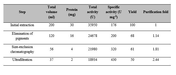
Table 1 shows the purification steps of GGP extract. After the elimination of pigments, GGP-specific activity was 200 U mg-1. An additional increase in the purification factor was observed after exclusion chromatography and ultrafiltration with a specific activity of 320 and 430 U mg-1 respectively, in a good concordance with the previous results of Centeno et al. [28].
CVs obtained from unmodified electrodes and GGP-modified electrodes in a PBS solution (10 mM pH 7.8 and a scan rate of 50 mV/s) give us information about the intrinsic redox process of GGP (Figure 1). A pair of well-defined redox peaks at 120 mV/10.5 mV are observed at the GGP modified SPGE, and no redox peaks are obtained at the SPGE unmodified electrode. Therefore, the redox peaks in Fig. 1A could be attributed to the heme Fe3+/2+ couple of the immobilized GGP. Similar behavior was observed for SPGOE modified with GGP (Fig. 1B), but the redox peak couple was obtained around 184 mV/59 mV for the anodic and cathodic peaks, respectively.
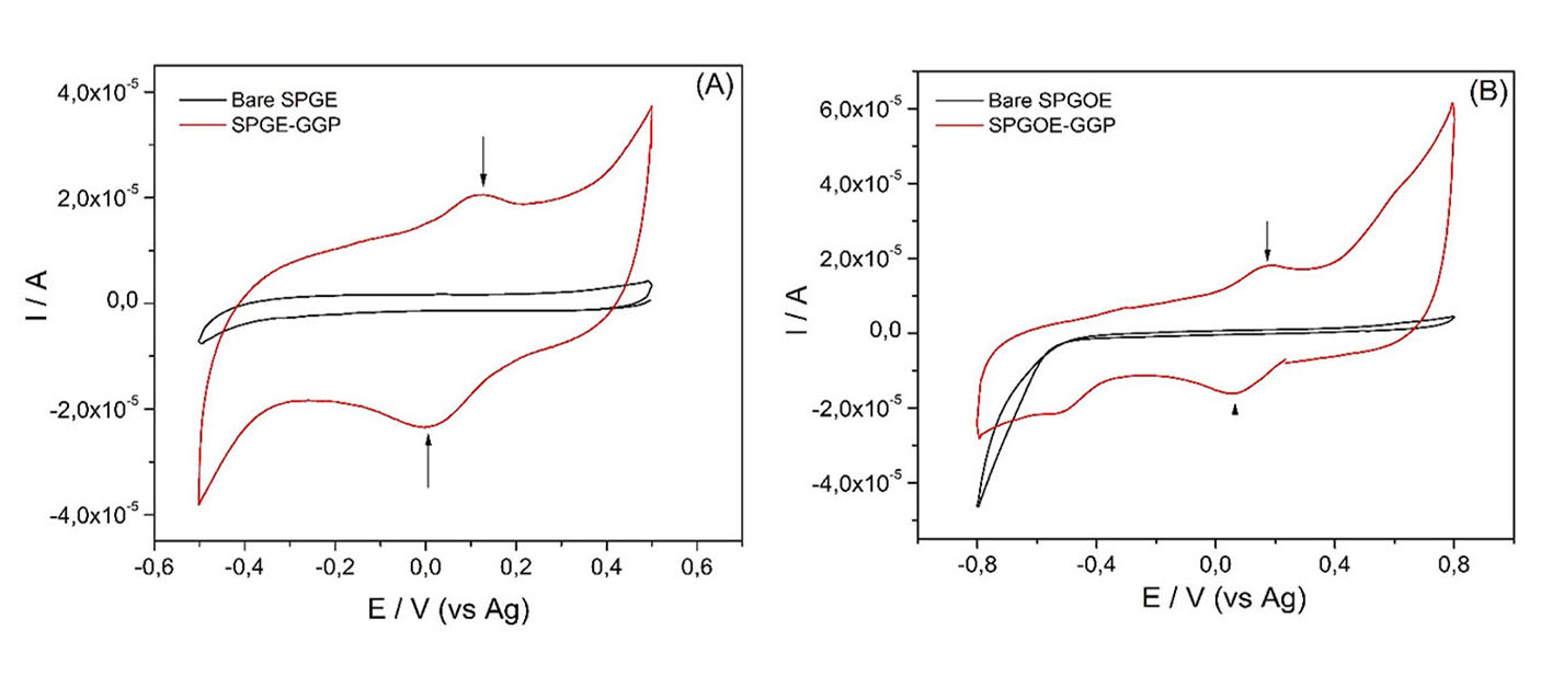
The formal potential (calculated as the average of the anodic and cathodic peak potentials) of GGP was ca. 65 mV and 121mV for SPGE and SPGOE electrodes, respectively. These dates are in good concordance with other heme Fe(III)/Fe(II) redox couple from plant PODs [29–31] and indicate that the graphene and graphene oxide surfaces create an excellent microenvironment for the immobilized enzyme and helps in better electron transfer between enzyme and electrode. Moreover, the peak-to-peak separation (∆Ep) was greater than 56/n mV (109 and 125 mV for SPGE and SPEGO at a scan rate of 50mV/s, respectively), suggesting the occurrence of a quasi-reversible direct electron transfer at the surface of screen-printed electrodes modified with GGP.
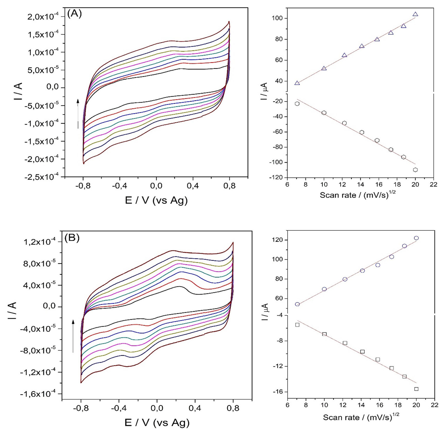
The effect of scan rate on the response of GGP immobilized on SPGE and SPGOE could provide information about reversibility of redox process. Figure 2 shows a linear dependence between scan rate root (ʋ) and anodic and cathodic peaks and the linear regression equations were ipa = 5.06ʋ+ 17.72 (R= 0.99), ipc = -0.75ʋ + 0.56 (R= 0.98) for SPGE-GGP and ipa = 4.85ʋ + 3.71 (R= 0.99), ipc = -6.59ʋ + 29.63 (R= 0.98), which indicated a typical surface-controlled quasi-reversible process of the electroactive species moving to the electrode surface [32].
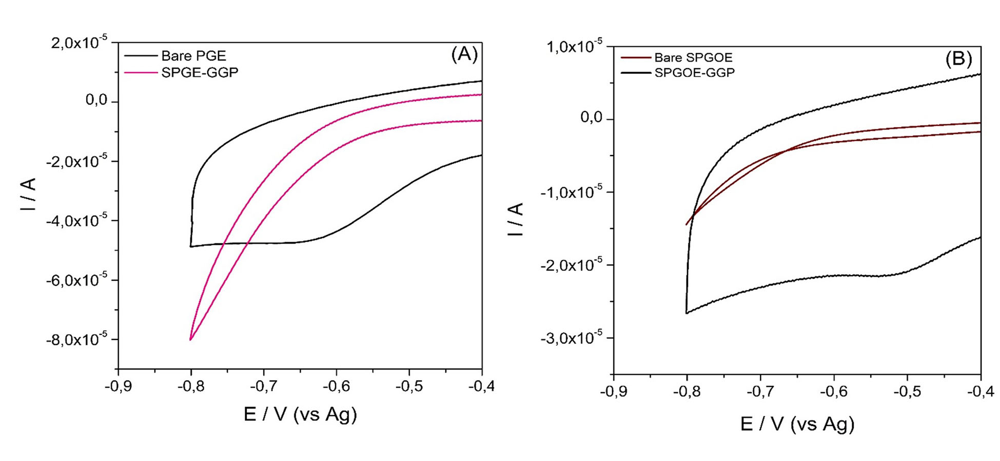
Electrocatalytic reduction of H2O2 on bare and modified screen-printed electrodes with peroxidase from guinea grass was monitored by a CV, as shown in Figure 3. There was no catalytic activity on bare electrodes in the presence of H2O2. On the other hand, when H2O2 was added into the electrochemical cell with SPGE and SPGOE modified electrodes, the voltammograms showed a high reduction current peak at -0.6 V and -0.5 V, respectively. This is attributed to the bio-electrocatalytic reduction of H2O2 by the presence of GGP acting as an efficient biocatalyst. Similar behavior was observed by Xu et al. [33] in reducing of H2O2 at -0.4 V by hemoglobin adsorbed in quantum dots electrodes. Moyo et al. obtained a redox potential of -0.5 V for H2O2 reduction by using HRP adsorbed onto a nano-biomaterial composite modified glassy carbon electrode [11].
It is also evident that the electrocatalytic efficacy for reducing H2O2 on SPGE-GGP (Figure 3A) is driving a higher reduction current than on SPGOE-GGP (Figure 3B). These results could demonstrate that SPGE displays better conductive properties and a large effective surface facilitating a fast electron transfer.
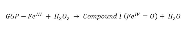
As with many other plant PODs and according to previous reports [34], the mechanism by which the GGP immobilized on the electrodes can reduce the hydrogen peroxide can be described by the following scheme:
 (1)
(1)
 (2)
(2)
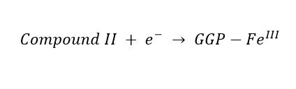 (3)
(3)After reacting with H2O2, the FeIII from heme group of GGP immobilized on the SPGE and SPGOE electrodes was oxidized to form compound I Equation 1, which contains a ferryl ion weakly spin-coupled to a porphyrin π-cation radical. Compound I obtain one electron from the electrode to form compound II, equation 2. Finally, compound II is reduced back to GGP native form by taking one electron from the surface electrode, as is shown in Equation 3[35,36].
The reduction of current dependence at different concentrations of H2O2 on the SPGE and SPGOE modified electrodes was evaluated using CVs measurements. Figure 4 displays different CVs in H2O2 concentrations between 0.1 to 4 mM for both electrodes.
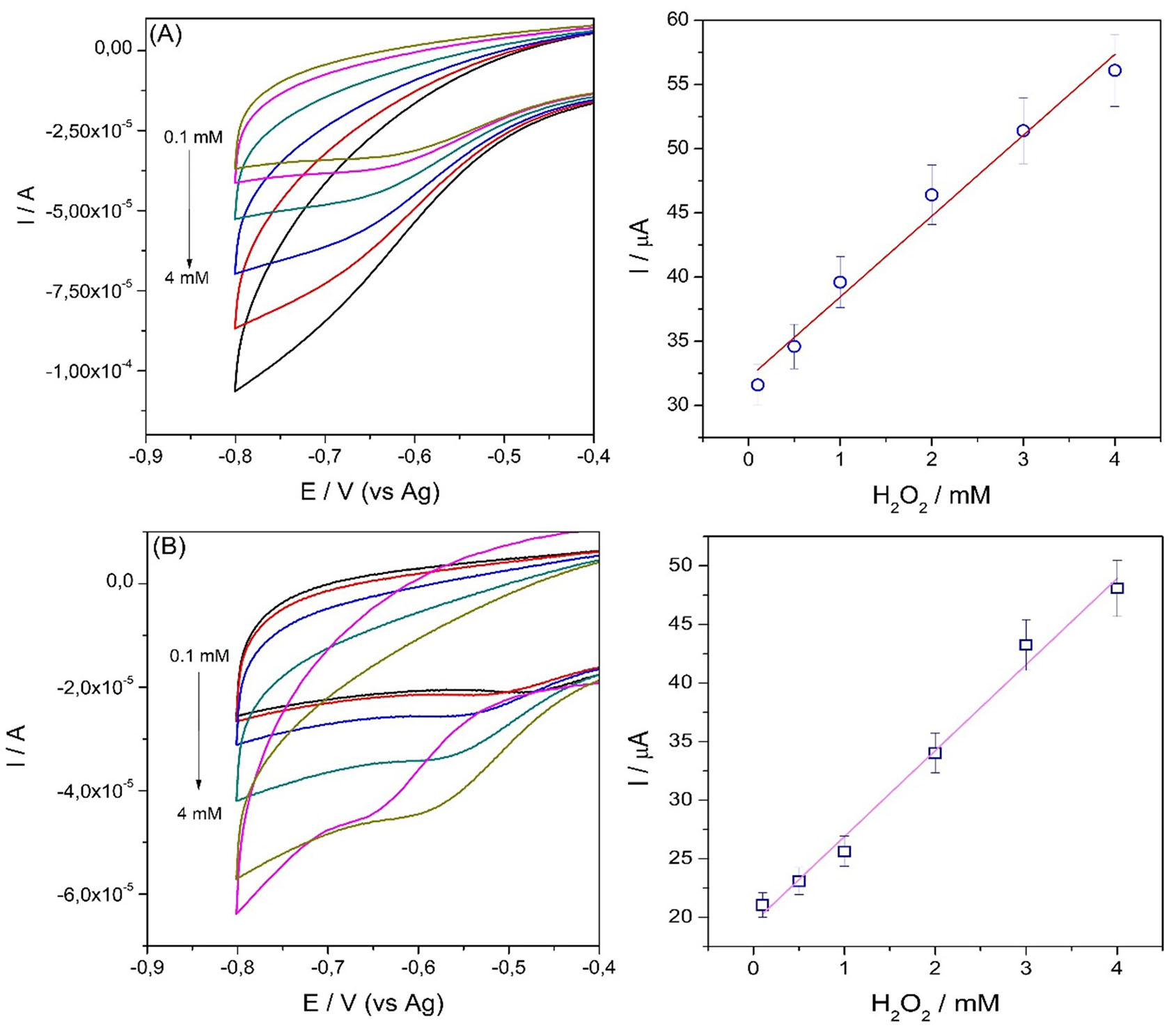
A linear response range of both modified electrodes towards different H2O2 concentrations was from 100 µM to 4 mM, and the linear equation regression where I = 6.29[H2O2] + 32.15 and I = 7.34[H2O2] + 19.54, with a correlation coefficient of 0.98 and 0.99 for SPGE-GGP and SPGOE-GGP, respectively.
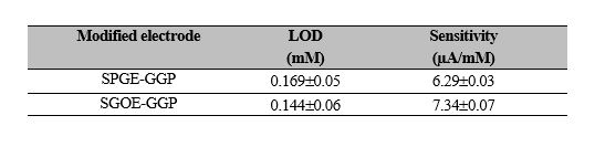
The limits of detection (LODs) were calculated based on the concept given by the International Union of Pure and Applied Chemistry (IUPAC) (CLOD = 3.3Sb/m, where Sb is the standard deviation of the blank and m the slope of the calibration curve) [33]. Sensitivities were calculated from the slope of the calibration curve obtained from current (µA) versus H2O2 concentration (mM) (Figure 4).
From Table 2, higher sensitivity was exhibited with the SPGE-GGP electrode, but when comparing the values of LOD, it can be observed that the lowest LOD was achieved with SPGOE-GGP.
According to these results, these two electrode configurations can be used to determine hydrogen peroxide in real samples.
Conclusions
This study is based on comparing bio-electrocatalytic reduction of H2O2 by screen-printed graphene and graphene oxide electrodes modified with guinea grass peroxidase (Panicum maximum). Both electrodes displayed a pair of well-redox peaks and an excellent efficiency reducing H2O2, although SPGE-GGP exhibited a higher electrocatalytic activity. Both electrodes exhibited a good performance towards H2O2 determination with a broader linearity range. SPGOE-GGP displayed a better LOD than SPGE, but SPGE-GGP obtained a higher sensitivity result. Further research with these novel electrodes integrating the high stable GGP will provide new bio-analytical tools for rapid and accurate detection of H2O2 in biomedical and environmental samples.
References
[1] L. G. Bach, M. L. N. Thi, N. T. Son, Q. B. Bui, H.-T. Nhac-Vu, and P. H. Ai-Le, “Mesoporous gold nanoparticles supported cobalt nanorods as a free-standing electrochemical sensor for sensitive hydrogen peroxide detection,” J. Electroanal. Chem., vol. 848, 2019. https://doi.org/10.1016/j.jelechem.2019.113359
[2] E. S. Abdel-Halim and S.S. Al-Deyab, “One-step bleaching process for cotton fabrics using activated hydrogen peroxide,” Carbohydr. Polym., vol. 92, pp. 1844–1849, 2013. https://doi.org/10.1016/j.carbpol.2012.11.045
[3] S. N. L. Lima, I. S. Ribeiro, M. A. Grisotto, E. S. Fernandes, V. Hass, R. R. J. Tavarez, S. C. S. Pinto, D. M. Lima, A. D. Loguercio and M. C. Bandeca, “Evaluation of several clinical parameters after bleaching with hydrogen peroxide at different concentrations: A randomized clinical trial,” J. Dent., vol. 68, pp. 91–97, 2018. https://doi.org/10.1016/j.jdent.2017.11.008
[4] Z. Li, H. Dou, Y. Fu and M. Qin, “Improving the hydrogen peroxide bleaching efficiency of aspen chemithermomechanical pulp by using chitosan,” Carbohydr. Polym., vol. 132, pp. 430–436, 2015. https://doi.org/10.1016/j.carbpol.2015.06.062
[5] F. Liu, L. Yang, X. Yin, X. Liu, L. Ge and F. Li, “A facile homogeneous electrochemical biosensing strategy based on displacement reaction for intracellular and extracellular hydrogen peroxide detection,” Biosens. Bioelectron., vol. 141, 2019. https://doi.org/10.1016/j.bios.2019.111446
[6] L. K. Ndungu, J. H. Steele, T. L. Hancock, R. D. Bartleson, E. C. Milbrandt, M. L. Parsons and H. Urakawa, “Hydrogen peroxide measurements in subtropical aquatic systems and their implications for cyanobacterial blooms,” Ecol. Eng., vol. 138, pp. 444–453, 2019. https://doi.org/10.1016/j.ecoleng.2019.07.011
[7] C. Peña-Bautista, M. Vento, M. Baquero and C. Cháfer-Pericás, “Lipid peroxidation in neurodegeneration,” Clin. Chim. Acta., vol. 497, pp. 178–188, 2019. https://doi.org/10.1016/j.cca.2019.07.037
[8] Y. Wen, F. Huo and C. Yin, “Organelle targetable fluorescent probes for hydrogen peroxide,” Chinese Chem. Lett., vol. 30, no. 10, 2019. https://doi.org/10.1021/ja100117u
[9] L. Zhang, P. Zhao, C. Yue, Z. Jin, Q. Liu, X. Du and Q. He, “Sustained release of bioactive hydrogen by Pd hydride nanoparticles overcomes Alzheimer’s disease,” Biomaterials, vol. 197, pp. 393–404, 2019. https://doi.org/10.1016/j.biomaterials.2019.01.037
[10] S. Farzana, V. Ganesh and S. Berchmans, “A Sensing Platform for Direct Electron Transfer Study of Horseradish Peroxidase,” J. Electrochem. Soc., vol. 160, no. 9, pp. H573–H580, 2013. https://iopscience.iop.org/article/10.1149/2.038309jes
[11] M. Moyo, J. O. Okonkwo and N. M. Agyei, “A novel hydrogen peroxide biosensor based on adsorption of horseradish peroxidase onto a nanobiomaterial composite modified glassy carbon electrode,” Electroanalysis, vol. 25, no. 8, pp. 1946–1954, 2013. https://doi.org/10.1002/elan.201300165
[12] G. S. Lai, H. L. Zhang and D. Y. Han, “Amperometric hydrogen peroxide biosensor based on the immobilization of horseradish peroxidase by carbon-coated iron nanoparticles in combination with chitosan and cross-linking of glutaraldehyde,” Microchim. Acta, vol. 165, pp. 159–165, 2009.
[13] W. Chen, S. Cai, Q. Q. Ren, W. Wen, and Y. Di. Zhao, “Recent advances in electrochemical sensing for hydrogen peroxide: A review,” Analyst, vol. 137, no. 1, pp. 49–58, 2012. https://doi.org/10.1039/C1AN15738H
[14] I. Y. Sakharov, “Palm tree peroxidases,” Biochem. Biokhimiia, vol. 69, pp. 823–829, 2004. https://doi.org/10.1023/b:biry.0000040213.91951.bc
[15] P. A. Uribe, C. C. Ortiz, D. A. Centeno, J. J. Castillo, S. I. Blanco, and J. A. Gutierrez, “Self-assembled Pt screen printed electrodes with a novel peroxidase Panicum maximum and zinc oxide nanoparticles for H.O. detection,” Colloids Surfaces A Physicochem. Eng. Asp., vol. 561, pp. 18–24, 2019. https://doi.org/10.1016/j.colsurfa.2018.10.051
[16] A. E. Orduz, J. A. Gutiérrez, S. I. Blanco, J. J. Castillo and J. C. Salcedo-Reyes, “Amperometric detection of triclosan with screen-printed carbon nanotube electrodes modified with Guinea Grass .Panicum maximum) peroxidase,” Universit. Scient., vol. 24, pp. 363–379, 2019. https://doi.org/10.11144/Javeriana.SC24-2.adot
[17] M. Li, S. Xu, M. Tang, L. Liu, F. Gao and Y. Wang, “Direct electrochemistry of horseradish peroxidase on graphene-modified electrode for electrocatalytic reduction towards H.O.,” Electrochim. Acta, vol. 56, no. 3, pp. 1144–1149, 2011. https://doi.org/10.1016/j.electacta.2010.10.034
[18] X. Bo, M. Zhou and L. Guo, “Electrochemical sensors and biosensors based on less aggregated graphene,” Biosens. Bioelectron, vol. 89, no. 1, pp. 167–186, 2017. https://doi.org/10.1016/j.bios.2016.05.002
[19] A. Bonanni, A. H. Loo and M. Pumera, “Graphene for impedimetric biosensing,” TrAC - Trends Anal. Chem., vol. 37, pp. 12–21, 2012. https://doi.org/10.1016/j.trac.2012.02.011
[20] B. Dinesh, V. Mani, R. Saraswathi and S.-M. Chen, “Direct electrochemistry of cytochrome c immobilized on a graphene oxide–carbon nanotube composite for picomolar detection of hydrogen peroxide,” RSC Adv., vol. 4, no. 54, 2014. https://doi.org/10.1039/C4RA02789B
[21] P. Olejnik, A. Świetlikowska, M. Gniadek and B. Palys, “Electrochemically Reduced Graphene Oxide on Electrochemically Roughened Gold as a Support for Horseradish Peroxidase,” J. Phys. Chem., vol. 118, pp. 129731-29738, 2014. https://doi.org/10.1021/jp507227z
[22] V. Vukojević, S. Djurdjić, M. Ognjanović, M. Fabian, A. Samphao, K. Kalcher and D.M. Stanković, “Enzymatic glucose biosensor based on manganese dioxide nanoparticles decorated on graphene nanoribbons,” J. Electroanal. Chem., vol. 823, pp. 610–616, 2018. https://doi.org/10.1016/j.jelechem.2018.07.013
[23] L. Li, H. Lu and L. Deng, “A sensitive NADH and ethanol biosensor based on graphene-Au nanorods nanocomposites,” Talanta, vol. 113, pp. 1–6, 2013. https://doi.org/10.1016/j.talanta.2013.03.074
[24] Y. Huang, Y. Xue. J. Zeng, S. Li, Z. Wang, C. Dong, G. Li, J. Liang, Z. Zhou. “Non-enzymatic electrochemical hydrogen peroxide biosensor based on reduction graphene oxide-persimmon tannin‑platinum nanocomposite”, Mater. Sci. Eng. C, vol. 92, pp. 590-598, 2018. https://doi.org/10.1016/j.msec.2018.07.021
[25] Z. Lu, L. Wu, X. Dai, Y. Wang, M. Sun, C. Zhou, H. Du and H. Rao. “Novel flexible bifunctional amperometric biosensor based on laser engraved porous graphene array electrodes: Highly sensitive electrochemical determination of hydrogen peroxide and glucose”. J. Hazard. Mater, vol. 402, pp. 123774, 2021. https://doi.org/10.1016/j.jhazmat.2020.123774
[26] S. Pandey, S. Sachan and S. Singh. “Electrochemically reduced graphene oxide modified with electrodeposited thionine and horseradish peroxidase for hydrogen peroxide sensing and inhibitive measurement of chromium”. Mat. Sci. Energy Techn, vol. 2, no. 3, pp. 676-686, 2019. https://doi.org/10.1016/j.mset.2019.08.001
[27] P. R. Matheus, J. M. Abad and V. M. Fernández, “Modification of gold surfaces for the oriented immobilization of recombinant form of horseradish peroxidase,” Rev. Téc. Ing. Univ. Zulia, vol. 30, no. 3, pp. 225–235, 2007. https://www.produccioncientificaluz.org/index.php/tecnica/article/view/6195
[28] D. A. Centeno, X. H. Solano and J. J. Castillo, “A new peroxidase from leaves of guinea grass (Panicum maximum): A potential biocatalyst to build amperometric biosensors,” Bioelectrochemistry, vol. 116, pp. 33-38, 2017.
[29] G. Battistuzzi, M. Bellei, C. A. Bortolotti and M. Sola, “Redox properties of heme peroxidases,” Arch. Biochem. Biophys., vol. 500, no. 1, pp. 21–36, 2010. https://doi.org/10.1016/j.abb.2010.03.002
[30] L. Lu, Y. Dong, J. Wang, Q. Li and, X. Wu, “Direct electrochemistry and bioelectrocatalysis of horseradish peroxidase entrapped in a self-supporting nanoporous gold electrode: A new strategy to improve the orientation of immobilized enzymes,” Anal. Methods., vol 7, no. 16, pp. 6686–6694, 2015. https://doi.org/10.1039/C5AY01333J
[31] Y.-X. Sun, J.-T. Zhang, S.-W. Huang and S.-F. Wang, “Hydrogen peroxide biosensor based on the bioelectrocatalysis of horseradish peroxidase incorporated in a new hydrogel film,” Sensors Actuators, B Chem., vol. 124, no. 2, pp. 494–500, 2007.
[32] Y. Wang, Z. Wang, Y. Rui and M. Li, “Horseradish peroxidase immobilization on carbon nanodots/CoFe layered double hydroxides: Direct electrochemistry and hydrogen peroxide sensing,” Biosens. Bioelectron., vol. 64, pp. 57–62, 2015. https://doi.org/10.1016/j.bios.2014.08.054
[33] Y. Xu, C. Hu and S. Hu, “A hydrogen peroxide biosensor based on direct electrochemistry of hemoglobin in Hb-Ag sol films,” Sensors Actuators, B. Chem., vol. 130, no. 2, pp. 816–822, 2008. https://doi.org/10.1016/j.snb.2007.10.048
[34] L. Shi, X. Liu, W. Niu, H. Li, S. Han, J. Chen and G. Xu, “Hydrogen peroxide biosensor based on direct electrochemistry of soybean peroxidase immobilized on single-walled carbon nanohorn modified electrode,” Biosens. Bioelectron., vol. 24, no. 5, pp. 1159–1163, 2009. https://doi.org/10.1016/j.bios.2008.07.001
[35] C. Ren, Y. Song, Z. Li and G. Zhu, “Hydrogen peroxide sensor based on horseradish peroxidase immobilized on a silver nanoparticles/cysteamine/gold electrode,” Anal. Bioanal. Chem., vol. 381, no. 6, pp. 1179–1185, 2005. https://doi.org/10.1007/s00216-004-3032-0
[36] F. Chekin, N. Leiva, J. B. Raoof, L. Gorton and L. Bülow, “Direct electrochemistry and bioelectrocatalysis of a class II non-symbiotic plant haemoglobin immobilised on screen-printed carbon electrodes,” Anal. Bioanal. Chem., vol. 398, no. 4, pp. 1643–1649, 2010. https://doi.org/10.1007/s00216-010-3800-y
Notes
*
Research paper (Artículo de investigación) - Article of scientific and tecnological investigation
Author notes
a Corresponding author. E-mail: jcasleon@uis.edu.co
Additional information
How to cite this article:: J. Castillo, P. A. Guarin-Guio, l. Ortiz, “Bio-electrocatalytic reduction of hydrogen peroxide by peroxidase from guinea grass (Panicum maximum) immobilized on graphene and graphene oxide screen-printed electrodes” Ing. Univ. vol. 26, 2022. https://doi.org/10.11144/javeriana.iued26.brhp
