
Palabras clave
biocompatibilidad
biomaterial
defecto óseo
hueso
periodoncia
quitosano
regeneración
regeneración ósea
tricálcico
Cómo citar

Resumen
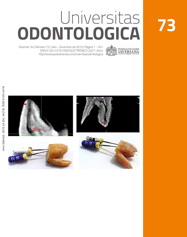
RESUMEN. Antecedentes: Se busca desarrollar injertos óseos biocompatibles capaces de regenerar defectos óseos de tamaño crítico. Objetivo: Evaluar la biocompatibilidad in vivo del fosfato tricálcico con quitosano (FTQ) en piel, músculo y hueso. Metodología: Se asignaron 15 ratas Wistar a grupos A (piel), B (músculo), C, D y E (defectos óseos de tamaño crítico). Se implantó FTQ en cada tejido. Como control se colocaron esponjas de colágeno adyacente a los sitios evaluados. Las ratas de los grupos A y B se sacrificaron a los 20 días, mientras que las de los grupos C, D y E se sacrificaron a los 20, 40 y 80 días respectivamente. Para confirmar la biocompatibilidad del FTQ, se evaluó la respuesta inflamatoria en términos de porcentaje: ninguna (0 %), leve (˂30 %), moderada (30-50 %) y alta (˃50 %), después de 20, 40 y 80 días en el tejido óseo. Resultados: No se encontró ulceración ni supuración en piel, músculo o hueso. Después de 80 días, el FTQ se observaba incorporado a tejido fibrótico y oseointegrado al hueso nativo. Conclusión: El FTQ fue biocompatible in vivo en piel, músculo y hueso.
ABSTRACT. Background: It is necessary to develop bone grafts capable to regenerate critical size bone defects. Objective: To evaluate the biocompatibility in vivo of tricalcium phosphate with chitosan (TPC) in skin, muscle, and bone. Methods: 15 Wistar rats were assigned to groups A (skin), B (muscle), C, D, and E (bone). TPC was placed in each tissue. In groups C-E, critical size bone defect was grafted with TPC and collagen sponge was placed adjacent to test sites as a control. Animals from groups A and B were sacrificed after 20 days, while groups C-E at days 45, 60, and 80. Inflammatory response was evaluated in all tissues. To assess biocompatibility, the percentage of cells was evaluated as none (0 %), low (˂ 30 %), moderate (30- 50 %), and high (˃50 %). Results: There were no signals of ulceration or suppuration in skin, muscle, and bone. After 80 days, TPC was incorporated into a fibrotic structure and osseointegrated to native bone. Conclusion: TPC was biocompatible with skin, muscle, and bone.
Mokbel N, Naaman N, Nohra J, Badawi N. Healing patterns of critical size bony defects in rats after grafting with bone substitutes soaked in recombinant human bone morphogenetic protein-2: histological and histometric evaluation. Br J Oral Maxillofac Surg. 2013; 51(6): 545-9.
Nasr HF, Aichelmann-Reidy ME, Yukna RA. Bone and bone substitutes. Periodontology 2000. 1999; 19: 74-86.
Misch CM. Autogenous bone: is it still the gold standard? Implant Dent. 2010; 19(5): 361.
Rossi AC, Freire AR, Prado FB, Caria PHF. Bone substitutes used in dentistry. Int J Odontostomatol. 2014; 8(2): 289-98.
Kim Y, Nowzari H, Rich SK. Risk of prion disease transmission through bovine-derived bone substitutes: a systematic review. Clin Implant Dent Related Res. 2013; 15(5): 645-53.
Sogal A, Tofe AJ. Risk assessment of bovine spongiform encephalopathy transmission through bone graft material derived from bovine bone used for dental applications. J Periodontol. 1999; 70(9): 1053-63.
Wenz B, Oesch B, Horst M. Analysis of the risk of transmitting bovine spongiform encephalopathy through bone grafts derived from bovine bone. Biomaterials. 2001; 22(12): 1599-606.
Lee K, Weir MD, Lippens E, Mehta M, Wang P, Duda GN, et al. Bone regeneration via novel macroporous CPC scaffolds in critical-sized cranial defects in rats. Dent Mater. 2014; 30(7): E199-E207.
Sculean A, Chapple ILC, Giannobile WV. Wound models for periodontal and bone regeneration: the role of biologic research. Periodontology 2000. 2015; 68(1): 7-20.
Yao CH, Liu BS, Hsu SH, Chen YS. Calvarial bone response to a tricalcium phosphate-genipin crosslinked gelatin composite. Biomaterials. 2005; 26(16): 3065-74.
Schlichting K, Dahne M, Weiler A. Biodegradable composite implants. Sports Med Arthrosc Rev. 2006; 14(3): 169-76.
Szabo G, Suba Z, Hrabak K, Barabas J, Nemeth Z. Autogenous bone versus beta-tricalcium phosphate graft alone for bilateral sinus elevations (2- and 3-dimensional computed tomographic, histologic, and histomorphometric evaluations): preliminary results. Int J Oral Maxillofac Implants. 2001; 16(5): 681-92.
Sato I, Akizuki T, Oda S, Tsuchioka H, Hayashi C, Takasaki AA, et al. Histological evaluation of alveolar ridge augmentation using injectable calcium phosphate bone cement in dogs. J Oral Rehab. 2009; 36(10): 762-9.
Kurashina K, Kurita H, Hirano M, Kotani A, Klein C, deGroot K. In vivo study of calcium phosphate cements: Implantation of an alpha-tricalcium phosphate dicalcium phosphate dibasic tetracalcium phosphate monoxide cement paste. Biomaterials. 1997; 18(7): 539-43.
Lee YM, Park YJ, Lee SJ, Ku Y, Han SB, Choi SM, et al. Tissue engineered bone formation using chitosan/tricalcium phosphate sponges. J Periodontol. 2000; 71(3): 410-7.
Muzzarelli RAA. Chitosan composites with inorganics, morphogenetic proteins and stem cells, for bone regeneration. Carbohydrate Polymers. 2011; 83(4): 1433-45.
Di Martino A, Sittinger M, Risbud MV. Chitosan: A versatile biopolymer for orthopaedic tissue-engineering. Biomaterials. 2005; 26(30): 5983-90.
Larsson KS. Screening-tests for systemic effects of dental materials. J Dent. 1994; 22: S12-S5.
Putters TF, Schortinghuis J, Vissink A, Raghoebar GM. A prospective study on the morbidity resulting from calvarial bone harvesting for intraoral reconstruction. Int J Oral Maxillofac Surg. 2015; 44(4): 513-7.
Arce Guerrero S, Valencia Llano C, Garzón-Alvarado DA. Obtención de un biocompuesto constituido por fosfato tricálcico y quitosana para ser usado como sustituto óseo en un modelo animal. Rev Cub Inv Biomed. 2012; 31(3): 268-77.
Ohura K, Bohner M, Hardouin P, Lemaitre J, Pasquier G, Flautre B. Resorption of, and bone formation from, new beta-tricalcium phosphate-monocalcium phosphate cements: An in vivo study. J Biomed Mater Res. 1996; 30(2): 193-200.
Fernandez T, Olave G, Valencia CH, Arce S, Quinn JMW, Thouas GA, et al. Effects of Calcium phosphate/chitosan composite on bone healing in rats: calcium phosphate induces osteon formation. Tissue Engineering Part A. 2014; 20(13-14): 1948-60.
Klokkevold PR, Vandemark L, Kenney EB, Bernard GW. Osteogenesis enhanced by chitosan (poly-N-acetyl glucosaminoglycan) in vitro. J Periodontol. 1996; 67(11): 1170-5.
Russel WMS, Burch RL. The principles of human experimental technique (1959). The global clearinghouse for information on alternatives to animal testing. Baltimore, MD: Johns Hopkins University; 2014. Disponible en: http://altweb.jhsph.edu/pubs/books/humane_exp/het-toc.
Shukla SK, Mishra AK, Arotiba OA, Mamba BB. Chitosan-based nanomaterials: A state-of-the-art review. Int J Biol Macromol. 2013; 59: 46-58.
Esta revista científica se encuentra registrada bajo la licencia Creative Commons Reconocimiento 4.0 Internacional. Por lo tanto, esta obra se puede reproducir, distribuir y comunicar públicamente en formato digital, siempre que se reconozca el nombre de los autores y a la Pontificia Universidad Javeriana. Se permite citar, adaptar, transformar, autoarchivar, republicar y crear a partir del material, para cualquier finalidad (incluso comercial), siempre que se reconozca adecuadamente la autoría, se proporcione un enlace a la obra original y se indique si se han realizado cambios. La Pontificia Universidad Javeriana no retiene los derechos sobre las obras publicadas y los contenidos son responsabilidad exclusiva de los autores, quienes conservan sus derechos morales, intelectuales, de privacidad y publicidad.
El aval sobre la intervención de la obra (revisión, corrección de estilo, traducción, diagramación) y su posterior divulgación se otorga mediante una licencia de uso y no a través de una cesión de derechos, lo que representa que la revista y la Pontificia Universidad Javeriana se eximen de cualquier responsabilidad que se pueda derivar de una mala práctica ética por parte de los autores. En consecuencia de la protección brindada por la licencia de uso, la revista no se encuentra en la obligación de publicar retractaciones o modificar la información ya publicada, a no ser que la errata surja del proceso de gestión editorial. La publicación de contenidos en esta revista no representa regalías para los contribuyentes.