
Cómo citar

Resumen
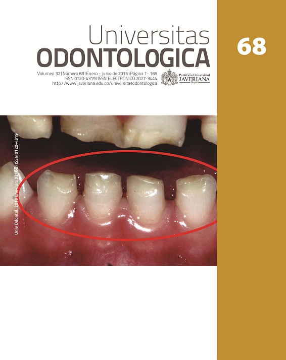
La gran diversidad de criterios usados para nombrar, definir, clasificar y medir los defectosen el desarrollo del esmalte (DDE) ha originado una amplia terminología ambiguae imprecisa que crea confusiones. Por ello, el propósito de este artículo es proponer laestandarización para beneficiar la salud pública, la investigación, la educación y la prácticaclínica. Para estandarizarlos, la Federación Dental Internacional hizo importantes avancesque plasmó en el índice DDE modificado (DDEm), que se toma como base con unas pocasprecisiones. Para medir los DDE en un primer tamizaje se sugiere usar el índice DDEm y sila prevalencia de algunos de los defectos es alta, se recomienda su medición específicacon índices de fluorosis o con los criterios para la hipomineralización incisomolar (HIM). Sinembargo, a futuro, unificar los métodos de medición sería ideal. También se sugiere repensarnombre y definición de la HIM.
The great diversity of criteria used to name, define, classify, and measure developmentaldefects of enamel (DDE) has resulted in a wide, ambiguous and imprecise terminology thatgenerates confusion. The purpose of this article is to propose standardization intended tobenefit public health, research, education, and clinical practice. In developing standards,the International Dental Federation has made great progress that resulted in the modifiedDDE Index (mDDE). Therefore, it is taken as a basis for this project with a few revisions. Tomeasure the presence of DDE in a first screening, it is recommended the use of the mDDEindex and if the prevalence of some of the defects is high, a specific measuring with fluorosisindices or criteria for the molar incisor hypomineralization (MIH) is recommended. However,unifying the methods for measurement would be advisable. It is also suggested to rethinkthe name and definition of the MIH.
Ellwood R, Fejerskov O, Cury JA, Clarkson B. Fluorides in caries control. En: Fejerskov O, Kidd E, editors. Dental caries: the disease and its clinical management. 2nd ed. Singapore: Blackwell Munksgaard; 2008. pp. 287-327.
Black GV, McKay FS. Mottled enamel: An endemic developmental imperfection of the teeth, heretofore unknown in the literature of dentistry. Dental Cosmos. 1916; 58: 129-56.
Dean HT. Classification of mottled enamel diagnosis. J Am Dent Assoc. 1934; 21: 1421-6.
Zimmermann ER. Fluoride and non-fluoride enamel opacities. Public Health Rep. 1954 Nov; 69(11): 1115-20.
Russell AL. The differential diagnosis of fluoride and non-fluoride enamel opacities. J Public Health Dent. 1961; 21; 143-6.
Clarkson J. Review of terminology, classifications, and indices of developmental defects of enamel. Adv Dent Res. 1989; 3(2): 104-9.
Clarkson J, O´Mullane D. A modified DDE index for use in epidemiological studies of enamel defects. J Dent Res. 1989 Mar; 68(3): 445-50.
International Dental Federation, Commission on Oral Health, Research and Epidemiology. A review of developmental defects of enamel index (DDE Index). Int Dent J. 1992; 42(6): 411-26.
World Health Organization. Oral health surveys. Basic methods. 4th ed. Geneva: WHO; 1997. pp. 34-6.
Weerheijm KL, Duggal M, Mejàre I, Papagiannoulis L, Koch G, Martens LC, Hallonsten A-L. Judgment criteria for Molar Incisor Hipomineralisation (HIM) in epidemiologic studies: a summary of the European meeting on MIH held in Athens. Eur J Paediatr Dent. 2003; (3): 110-3.
Fejerskov O, Nyvad B, Kidd E. Pathology of dental caries. En: Fejerskov O, Kidd E, editors. Dental caries: the disease and its clinical management. 2nd ed. Singapore: Blackwell Munksgaard; 2008. pp. 21-48.
Simmer JP, Papagerakis P, Smith CE, Fisher DC, Rountrey AN, Zheng L, Hu JC. Regulation of dental enamel shape and hardness. J Dent Res. 2010 Oct; 89(10): 1024-38.
Mahoney P. Two dimensional patterns of human enamel thickness on deciduous (dm1, dm2) and permanent first (M1) mandibular molars. Arch Oral Biol. 2010 Feb; 55(2): 115-26.
Tafforeau P, Smith TM. Nondestructive imaging of hominoid dental microstructure using phase contrast X-ray synchrotron microtomography. J Hum Evol. 2008; 54: 272-78.
Mahoney P. Intraspecific variation in M1 enamel development in modern humans: implications for human evolution. J Hum Evol. 2008 Jul; 55(1):131-47.
Richards A, Kragstrup J, Josephsen K, Fejerskov O. Dental fluorosis developed in post-secretory enamel. J Dent Res. 1986 Dec; 65(12): 1406-9.
Aoba T, Fejerskov O. Dental fluorosis: chemistry and biology. Crit Rev Oral Biol Med. 2002 Mar; 13(2): 155-70.
Farah R, Drummond B, Swain M, Williams S. Linking the clinical presentation of molar incisor hypomineralization to its mineral density. Int J Paediatr Dent. 2010 Sep; 20(5): 353-60.
Da Costa-Silva CM, Ambrosano GMB, Jeremias F, De Souza JF, Mialhe FL. Increase in severity of molar–incisor hypomineralization and its relationship with the colour of enamel opacity: a prospective cohort study. Int J Paediatr Dent. 2011 Sep; 21(5): 333-41.
Addy M, Moran J. Mechanisms of stain formation on teeth, in particular associated with metal ions and antiseptics. Adv Dent Res. 1995 Dec; 9(4): 450-6.
Trancho GJ, Robledo B. Patología oral: hipoplasia del esmalte dentario. Madrid: Departamento de Biología Animal (Antropología), Facultad de Biología, Universidad Complutense de Madrid; 2000.
Finchman AG, Moradian-Oldak J, Simmer JP. The structural biology of the developing dental enamel matrix. J Struct Biol. 1999 Jun 30; 126(3): 270-99.
Antoine D, Hillson S, Dean MC. The developmental clock of dental enamel: a test for the periodicity of prism cross-striations in modern humans and an evaluation of the most likely sources of error in histological studies of this kind. J Anat. 2009 Jan; 214(1): 44-55.
Woźniak K, Łagocka R, Lipski M, Tomasik M, Buczkowska-Radli´nska J, Chlubek D. Changes in developmental defects of dental enamel within the space of centuries. Durham Anthropol [internet]. 2005; 12(2-3). Avalaible from: http://www.dur.ac.uk/anthropology.journal/vol12/iss2-3/wozniak/wozniak.html.
Griffin RC, Donlon D. Patterns in dental enamel hypoplasia by sex and age at death in two archaeological populations. Arch Oral Biol. 2009 Dec; 54(1): 93-100.
Guatelli-Steinberg D. What can developmental defects of enamel reveal about physiological stress in nonhuman primates? Evol Anthropol. 2001; 10(4): 138-51.
Atar M, Körperich EJ. Systemic disorders and their influence on the development of dental hard tissues: a literature review. J Dent. 2010 Apr; 38(4): 296-306.
Moreno S, León M, Marín L, Moreno F. Comportamiento in vitro de los tejidos dentales y de algunos materiales de obturación dental sometidos a altas temperaturas con fines forenses. Colomb Med. 2008 Ene-Mar; 39(1): 28-46.
Seow WK, Clinical diagnosis of enamel defects: pitfalls and practical guidelines. Int Dent J. 1997 Jun; 47(3): 173-82.
Machiulskiene V, Baelum V, Fejerskov O, Nyvad B. Prevalence and extent of dental caries, dental fluorosis and developmental enamel defects in Lithuanian teenage populations with different fluoride exposures. Eur J Oral Sci 2009 Apr; 117(2): 154-60.
Segovia A, Estrella R, Medina CE, Maupomé G. Severidad de caries y factores asociados en preescolares de 3-6 años de edad en Campeche, México. Rev Salud Pública. 2005; 7(1): 756-69.
Vallejos AA, Medina CE, Casanova JF, Maupomé G, Casanova AJ, Minaya M. Defectos del esmalte, caries en dentición primaria, fuentes de fluoruro y su relación con caries en dientes permanentes. Gac Sanit. 2007 May-Jun; 21(3): 227-34.
Lygidakis NA, Dimou G, Briseniou E. Molar-incisor-hypomineralisation (MIH). Retrospective clinical study in Greek children: I. Prevalence and defect characteristics. Eur Arch Paediatr Dent. 2008 Dec; 9(4): 200-6.
Rodd HD, Boissonade FM, Day PF. Pulpal status of hypomineralized permanent molars. Pediatr Dent. 2007 Nov-Dec; 29(6): 514-20.
Jälevik B, Klingberg GA. Dental treatment, dental fear and behaviour management problems in children with severe enamel hypomineralization of their permanent first molars. Int J Paediatr Dent. 2002 Jan; 12(1): 24-32.
Marshman Z, Gibson B, Robinson PG. The impact of developmental defects of enamel on Young people in the UK. Community Dent Oral Epidemiol. 2009 Feb; 37(1): 45-57.
Jälevik B, Klingberg GA. Treatment outcomes and dental anxiety in 18-year-olds with MIH, comparisons with healthy controls - a longitudinal study. Int J Paediatr Dent. 2012 Mar; 22(2):85-91.
International Caries Detection and Assessment System Coordinating Committee. Rationale and Evidence for the International Caries Detection and Assessment System (ICDAS II) [internet]. 2011. Available from: http://www.icdas.org/downloads.
Møller IJ. Fluorides and dental fluorosis. International Dent J.1982; 32(2): 135-47.
Brook AH, Elcock C, Hallonstein AL, Poulsen S, Andreassen J, Koch G, Yeung CA, Dosanjh T. The development of a new index to measure enamel defects. In: Brook AH, editor. Dental morphology. Sheffield, UK: Sheffield Academic Press; 2001. pp. 59-66.
International Dental Federation, Commission on Oral Health, Research and Epidemiology. An epidemiological index of developmental defects of dental enamel (DDE Index). Int Dent J. 1982 Jun; 32(2): 159-67.
World Health Organization. Oral health surveys. Basics methods. 3rd ed. Geneva: WHO; 1987.
Thylstrup A, Fejerskov O. Clinical appearance and surface distribution of dental fluorosis in permanent teeth in relation to histological changes. Community Dent Oral Epidemiol. 1978 Nov; 6(6): 315-28.
Horowitz HS, Driscoll WS, Meyers RJ, Heifetz SB, Kigman A. A New Method of Assessing the Prevalence of Fluorosis - The Tooth Surface Index of Fluorosis. J Am Dent Assoc. 1984 Jul; 109(1): 37-41.
Dean HT. The Investigation of physiological effects by the epidemiological method. En: Moulton FR, editor. Fluorine and dental health. Washington, DC: American Association for the Advancement of Science; 1942.
Pendrys DG. The fluorosis risk index: a method for investigating risk factors. J Public Health Dent. 1990 Fall; 50(5): 291-8.
Rozier RG. Epidemiologic indices for measuring the clinical manifestations of dental fluorosis: overview and critique. Adv Dent Res. 1994 Jun; 8(1): 39-55.
Jalevik B. Prevalence and diagnosis of molar-incisor-hypomineralisation (MIH): A systematic review. Eur Arch Paediatr Dent. 2010 Apr; 11 (2): 59-64.
Arrow P. Prevalence of developmental enamel defects of the permanent molars among school children in Western Australia. Aus Dent J. 2008; 53: 250-9.
Balmer RC, Laskey D, Mahoney E, Toumba KJ. Prevalence of enamel defects and MIH in non-fluoridated and fluoridated communities. Eur J Paediatr Dent. 2005 Dec; 5(4): 209-12.
Dietrich G, Sperling S, Hetzer G. Molar Incisor Hypomineralisation in a group of children and adolescents living in Dresden (Germany). Eur J Paediatr Dent 2003 Sep; 4(3): 133-7.
Calderara PC, Gerthoux PM, Mocarelli P, et al. The prevalence of Molar Incisor Hypomineralisation (MIH) in a group of Italian school children. Eur J Paediatr Dent. 2005 Jun; 6(2): 79-83.
Fleita D, Ali A, Alaluusua S. Molar-incisor hypomineralisation (MIH) in a group of school-aged children in Benghazi, Libya. Eur Arch Paediatr Dent. 2006 Jun; 7(2): 92-5.
Esta revista científica se encuentra registrada bajo la licencia Creative Commons Reconocimiento 4.0 Internacional. Por lo tanto, esta obra se puede reproducir, distribuir y comunicar públicamente en formato digital, siempre que se reconozca el nombre de los autores y a la Pontificia Universidad Javeriana. Se permite citar, adaptar, transformar, autoarchivar, republicar y crear a partir del material, para cualquier finalidad (incluso comercial), siempre que se reconozca adecuadamente la autoría, se proporcione un enlace a la obra original y se indique si se han realizado cambios. La Pontificia Universidad Javeriana no retiene los derechos sobre las obras publicadas y los contenidos son responsabilidad exclusiva de los autores, quienes conservan sus derechos morales, intelectuales, de privacidad y publicidad.
El aval sobre la intervención de la obra (revisión, corrección de estilo, traducción, diagramación) y su posterior divulgación se otorga mediante una licencia de uso y no a través de una cesión de derechos, lo que representa que la revista y la Pontificia Universidad Javeriana se eximen de cualquier responsabilidad que se pueda derivar de una mala práctica ética por parte de los autores. En consecuencia de la protección brindada por la licencia de uso, la revista no se encuentra en la obligación de publicar retractaciones o modificar la información ya publicada, a no ser que la errata surja del proceso de gestión editorial. La publicación de contenidos en esta revista no representa regalías para los contribuyentes.