
Keywords
cone-beam computed tomography
dentistry
mandibular condyle
sleep bruxism
temporomandibular joint
temporomandibular disorders
How to Cite

Abstract
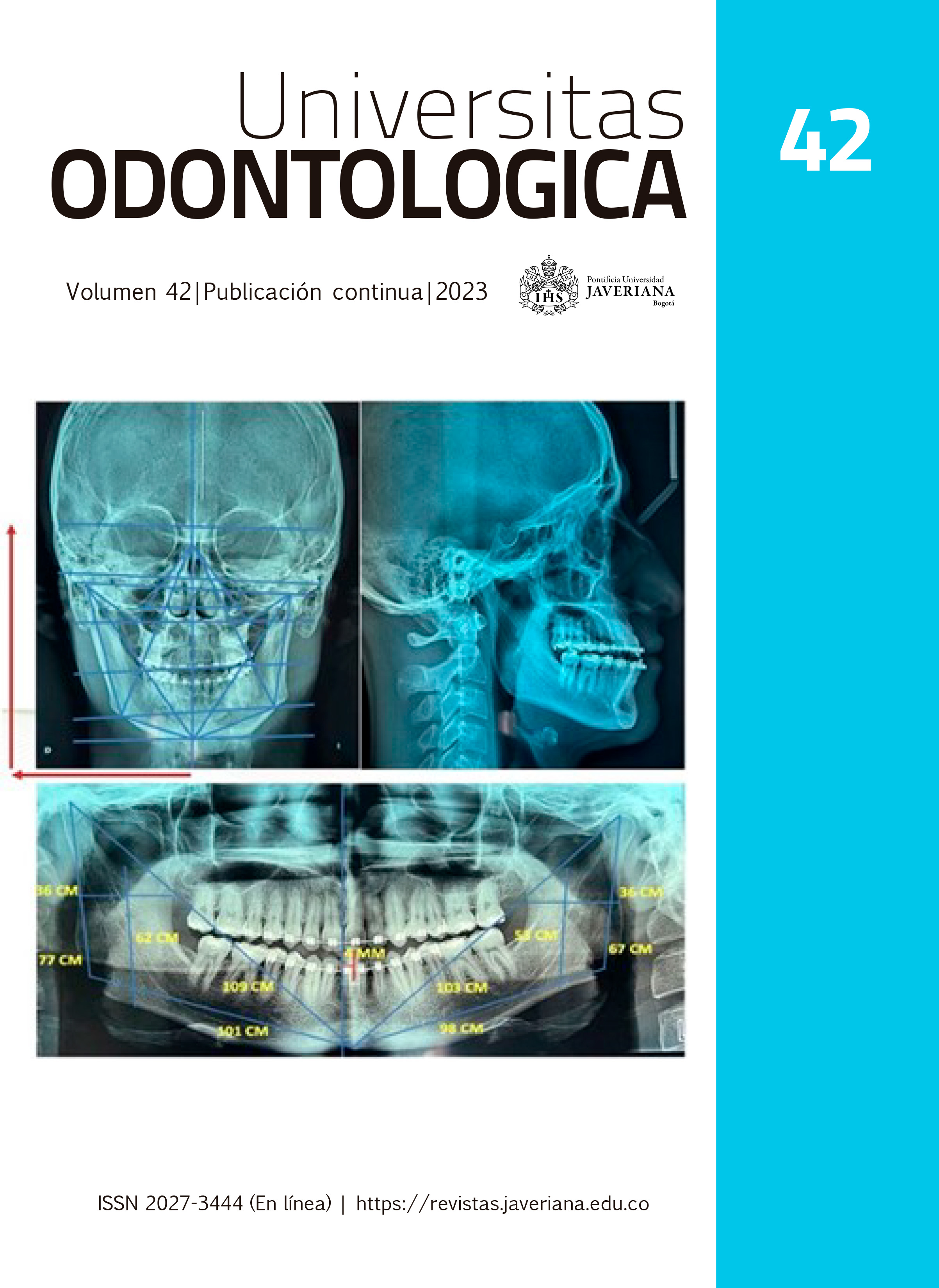
Objective: To establish the relationship between the condylar position of the temporomandibular joint (TMJ) using cone beam computed tomography (CBCT) with the dental wear patterns determined with the Bruxchecker®, in subjects with a clinical diagnosis of sleep bruxism. Method: Observational, descriptive, cross-sectional study in 45 patients with clinical diagnosis of sleep bruxism, according to the bruxism diagnostic criteria of the AASM (American Academy of Sleep Medicine), with an average age of 34.9 ± 8.6. The CBCT were analyzed, measuring the spaces of the TMJ in the sagittal plane: anterior, superior and posterior (SA, SS, SP) and in the coronal plane: lateral, superior and medial (Cl, CS, CM). The pattern of dental wear was classified with the Bruxchecker® by dividing the subjects into: Group 1, canine pattern (CP) with 19 subjects and Group 2, molar pattern (MP) with 26 subjects. Results: No significant differences were observed between the measurements of the joint spaces of each group nor in the comparison between the PC vs PM patterns. The sagittal position of the condyle posterior/anterior(P/A) was posterior and the coronal medial/lateral (M/L) was medial for the two groups, when evaluating the ratios. Significant difference (P <0.00) was found in right pattern CP and MP, when estimating the difference between averages of medial and lateral spaces. Conclusions: The position of the condyle was found posterior and medial in the articular fossa, in the right CP and in the left MP. A significant right lateral condyle position was evidenced in PC and PM. These findings indicate an altered condylar position in subjects with bruxism, which can be associated with joint distraction, possibly generated by constant eccentric mandibular movements during bruxism.

This work is licensed under a Creative Commons Attribution 4.0 International License.
Copyright (c) 2024 Ivonne Casalins Rolong, Judith Patricia Barrera Chaparro, Karen Julieth Bonilla, Sofía Hernández Vargas