
Palabras clave
articulación temporomandibular
BruxChecker®
bruxismo del sueño
odontología
tomografía computarizada de haz cónico
trastornos temporomandibulares
BruxChecker®
bruxismo del sueño
odontología
tomografía computarizada de haz cónico
trastornos temporomandibulares
Cómo citar
1.
CASALINS ROLONG I, Barrera Chaparro JP, Bonilla KJ, Hernández Vargas S. RELACIÓN ENTRE LA POSICIÓN CONDILAR DE LA ARTICULACIÓN TEMPOROMANDIBULAR Y EL PATRÓN DE DESGASTE DENTAL, EN SUJETOS CON DIAGNÓSTICO CLÍNICO DE BRUXISMO DEL SUEÑO . Univ Odontol [Internet]. 2023 Dec. 31 [cited 2025 Dec. 6];42. Available from: https://revistas.javeriana.edu.co/index.php/revUnivOdontologica/article/view/38025
Almetrics
Dimensions
Google Scholar

Resumen
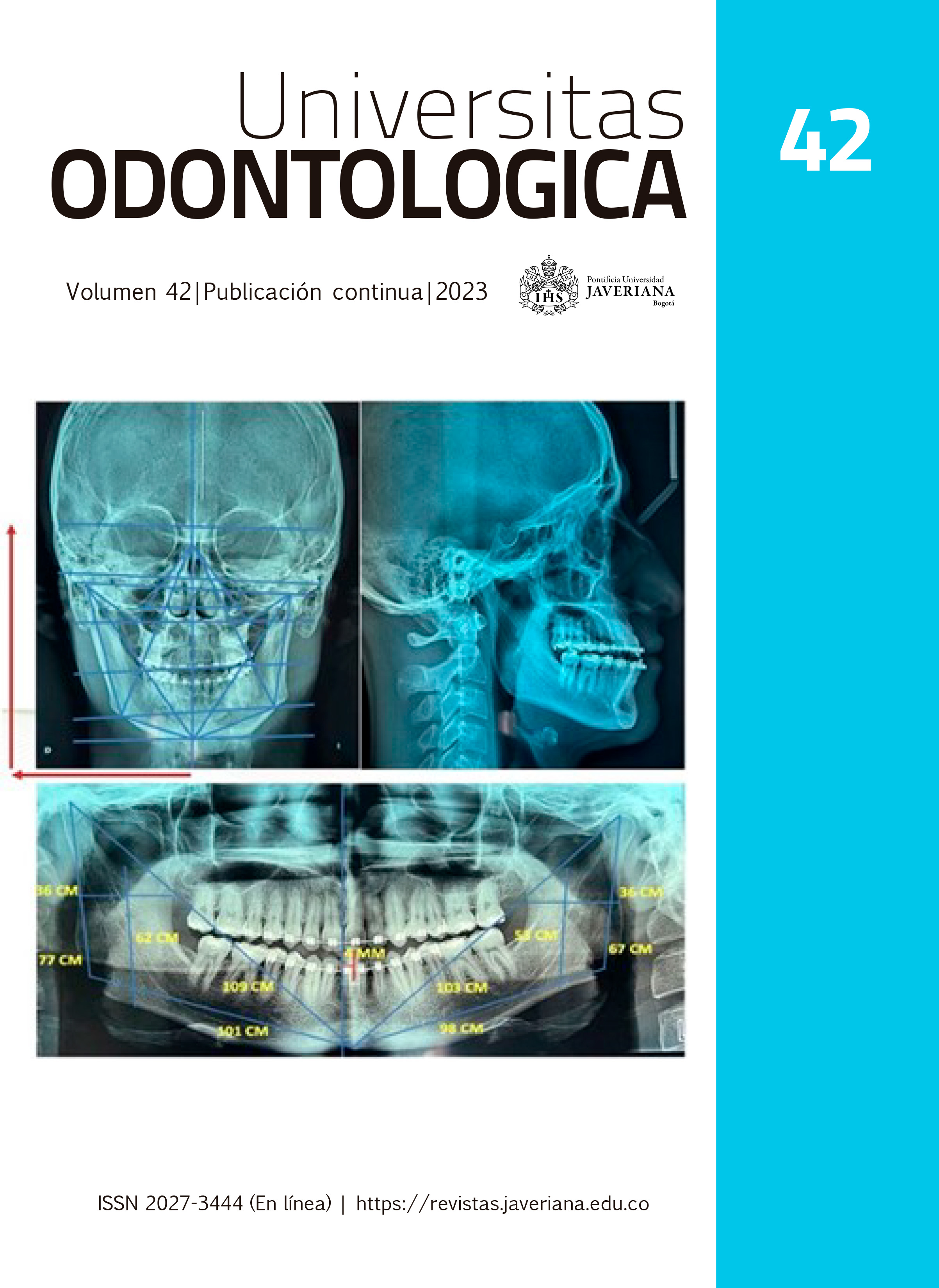
Objetivo: Establecer la relación entre la posición condilar de la articulación temporomandibular (ATM) utilizando tomografías computarizadas de haz cónico (TCHC) con los patrones de desgaste dental determinados con el Bruxchecker®, en sujetos con diagnóstico clínico de bruxismo del sueño. Método: Estudio observacional, descriptivo, transversal, en 45 pacientes con diagnóstico clínico de bruxismo del sueño, según los criterios de diagnóstico del bruxismo de la AASM (American Academy of Sleep Medicine), con edad promedio de 34,9 ± 8,6. Se analizaron las TCHC, midiendo los espacios de la ATM en el plano sagital: anterior, superior y posterior (SA,SS,SP) y en el plano coronal: lateral, superior y medial (CL,CS,CM). Se clasificó el patrón de desgaste dental con el Bruxchecker® dividiendo los sujetos en: Grupo 1, patrón canino (PC) con 19 sujetos y Grupo 2, patrón molar (PM) con 26 sujetos. Resultados: No se observaron diferencias significativas entre las medidas de los espacios articulares de cada grupo ni en la comparación entre los patrones PC vs PM. La posición sagital del cóndilo posterior/anterior (P/A) fue posterior y la coronal medial/lateral (M/L) fue medial para los dos grupos, al evaluar los ratios. Se encontró diferencia significativa (P<0,00) en los patrones PC y PM del lado derecho, al estimar la diferencia entre promedios de espacios medial y lateral. Conclusiones: La posición del cóndilo se encontró posterior y medial en la fosa articular, en los patrones PC derecho y PM izquierdo. Se evidenció una posición condilar lateral derecha significativa, en los patrones PC y PM. Estos hallazgos indican una posición condilar alterada en los sujetos con bruxismo, que se puede asociar con una distracción articular, generada posiblemente por los movimientos excéntricos mandibulares constantes durante la actividad de bruxismo.
Esta obra está bajo una licencia internacional Creative Commons Atribución 4.0.
Derechos de autor 2024 Ivonne Casalins Rolong, Judith Patricia Barrera Chaparro, Karen Julieth Bonilla, Sofía Hernández Vargas