
Cómo citar

Resumen
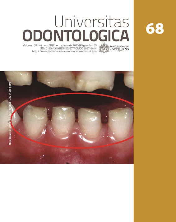
Antecedentes: El esmalte dental, el tejido más duro del cuerpo, puede ser destruido por la caries, una enfermedad infecciosa y transmisible causada por bacterias. Cuando trastornos genéticos y hereditarios, como la amelogénesis imperfecta (AI), afectan la formación del esmalte, este se hace más vulnerable a sufrir dicha patología. Objetivo: Describir el tipo de caries presente en nueve individuos de ocho familias colombianas con diferentes fenotipos de AI. Métodos: Se realizó análisis clínico, de radiografías periapicales y panorámicas, y de fotografías. Se utilizó la clasificación de Witkop para los fenotipos de AI y la del Sistema Internacional de Valoración y Detección de Caries (ICDAS) para caries dental. Resultados: Los pacientes con AI, fenotipo hipoplásico, presentaron registros ICDAS 5 y 6. En algunos pacientes con AI, fenotipo hipocalcificado, se encontraron registros ICDAS 3 y 4 y desgastes dentales graves; mientras que en los pacientes con fenotipo hipomadurativo los registros ICDAS estuvieron entre 1 y 3. Conclusiones: El gran número de registros ICDAS 5 y 6 en pacientes con AI fenotipo hipoplásico parece estar relacionado con factores como rugosidad, sensibilidad y susceptibilidad a la fractura del esmalte. En pacientes con AI fenotipo hipocalcificado, los registros 3 y 4 posiblemente fueron causados por el esmalte poco mineralizado y poroso. Los desgastes parecen actuar como una forma de microabrasión que elimina el tejido cariado. En el fenotipo hipomadurativo se presentaron los registros más bajos, lo que parece deberse al gran contenido de proteína anormal que actúa como un protector de la disolución del esmalte por los ácidos.
Background: Dental enamel, the human hardest tissue, can be destroyed by tooth decay, an infectious and transmissible disease caused by bacteria. When genetic or heredity disorders such as imperfect amelogenesis (AI) affect the enamel formation, it makes it more vulnerable to the onset of caries or tooth decay. Objective: Describe the type of tooth decay present in nine individuals from eight Colombian families with different AI phenotypes. Methods: Clinical, radiographic (periapical and panoramic radiographs), and photographic analysis were performed. AI phenotypes were classified through Witkop’s index and so was ICDAS for dental caries. Results: Patients with hypoplastic phenotype of AI had ICDAS scores of 5 and 6. In some patients with hypocalcified phenotype of AI ICDAS 3 and 4 and severe dental wear were found. In addition, patients with hypomadurative phenotype had ICDAS scores 1-3. Conclusions: The larger number of ICDAS scores 5 and 6 in patients with hypoplastic AI appears to be due to factors such as roughness, sensitivity, and susceptibility to enamel fracture. In patients with hypocalcified AI ICDAS 3 and 4 were possibly caused by the porous and poorly mineralized enamel. Wear seems to act as a means of microabrasion to eliminate the carious tissue. The lowest ICDAS scores present in hypomadurative AI seem to be due to the high content of abnormal phenotype protein that acts as a dissolution protector of the enamel by acids.
Robinson C, Shore RC, Bonass WA, Brookes SJ, Boteva E, Kirkham J. Identification of human serum albumin in human caries lesions of enamel: the role of putative inhibitors of remineralization. Caries Res. 1998; (32): 193-9.
Fejerskov O, Kidd EAM, Nyvad B, et al. Defining the disease: an introduction. In: Fejerskov O, Kidd E, editors. Dental caries: the disease and its clinical management. 2nd edition. Oxford: Blackwell Munksgaard; 2008. pp. 4-6.
Tenovuo J. Antimicrobial function of human saliva–how important is it for oral health? Acta Odontol Scand. 1998; Oct 56(5): 250-6.
Kawasaki K, Tanaka Y, Tkagi O. Crystallographic analysis of demineralized human enamel treated. Arch Oral Biol. 2000 Sep; 45(9): 797-804.
Fejerskov O. Concepts of dental caries and their consequences for understanding the disease. Community Dent Oral Epidemiol. 1997 Feb; 25(1): 5-12.
Zeichner-David M. Is there more to enamel matrix proteins than biomineralization? Matrix Biol. 2001 Sep; 20(5-6): 307-16.
Robinson C, Brookes SJ, Shore RC, Kirkham J. The developing enamel matrix: nature and function. Eur J Oral Sci. 1998 Jan; 106(Suppl): 282-91.
Robinson C, Kirkham J, Brookes SJ, Bonass WA, Shore RC. The chemistry of enamel development. Int J Dev Biol. 1995 Feb; 39(1): 145-52.
Robinson C, Brookes SJ, Bonass WA, Shore RC, Kirkham J. Enamel maturation. Ciba Found Symp. 1997; 205: 156-70; discussion 170-4.
Moradian-Oldak J, Paine ML. Mammalian dental enamel. In: Sigel HSA, Sigel RKO, editors. Biomineralization: From nature to application. Chichester (UK): John Wiley & Sons; 2008. pp. 507-46.
Witkop CJ Jr, Sauk JJ Jr. Heritable defects of enamel. In: Stewart RE, Prescott GH, editors. Oral facial genetics. St. Louis, Missouri: Mosby; 1976. pp. 151-226.
Witkop CJ. Amelogenesis imperfecta, dentinogenesis imperfecta and dentin dysplasia revisited: problems in classification. J Oral Pathol. 1988 Nov; 17(9-10): 547-53.
Gutiérrez SJ, Torres DM, Briceño I, Gómez A, Baquero E. Clinical and molecular analysis of the enamelin gene ENAM in Colombian families with autosomal dominant amelogenesis imperfecta. Genet Mol Biol. 2012 Jul; 35(3): 557-66.
Ismail Al, Tellez, Sohn W, Sen A. The International Caries Detection and Assessment System (ICDAS): an integrated system for measuring dental caries. Community Dent Oral Epidemiol. 2007 Jun; 35(3): 170-8.
Mahoney EK, Rohanizadeh R, Ismail FS, Kilpatri'ck NM, Swain MV. Mechanical properties and microstructure of hypomineralised enamel of permanent teeth. Biomaterials. 2004 Sep; 25(20): 5091-100.
Mandel ID. Nature versus nurture in dental caries. J Am Dent Assoc. 1994 Oct; 125(10): 1345-51.
Seymen F, Kiziltan B. Amelogenesis imperfecta: a scanning electron microscopic and histopathologic study. J Clin Pediatr Dent. 2002 Summer; 26(4): 327-35.
Boyde A. A 3-D model of enamel development at the scale of one inch to the micron. Adv Dent Res. 1987 Dec; 1(2): 135-40.
Mahoney E, Ismail FS, Kilpatrick N, Swain M. Mechanical properties across hypomineralized/hypoplastic enamel of first permanent molar teeth. Eur J Oral Sci. 2004 Dec; 112(6): 497-502.
LeGeros RZ. Calcium phosphates in enamel, dentin and bone. In: Myers HM, editor. Calcium phosphates in oral biology and medicine. Basel: Karger; 1991. pp. 108-29.
Angker L, Nokolds C, Swin M, Kilpatrick N. Quantitative analysis of the mineral content of sound and carious primary dentine using BSE imaging. Arch Oral Biol. 2004 Feb; 49(2): 99-107.
Weerheijm KL, Jalevik B, Alaluusua S. Molar- incisor hypomineralisation. Caries Res. 2001 Sep-Oct; 35(5): 390-1.
Leppaniemi A, Lukinmaa PL, Alaluusua S. Non fluoride hypomineralizations in the permanent first molars and their impact on the treatment need. Caries Res. 2001 Jan-Feb; 35(1): 36-40.
Croll TP. Hastening the enamel microabrasion procedure. Eliminating defects, cutting treatment time. J Am Dent Assoc. 1993 Apr 1; 124: 487-90.
Robinson C, Kirkham J. Enamel matrix components, alterations during development and possible interactions with the mineral phase. Tooth Enamel IV. Amsterdam: Elsevier; 1984. pp. 261-5.
Kubota K, Lee DH, Tsuchiya M, Young CS, Everett ET, Martinez-Mier EA, et al. Fluoride induces endoplasmic reticulum stress in ameloblasts responsible for dental enamel formation. J Biol Chem. 2005 Jun 17; 280(24): 23194-202.
Smith CE. Cellular and chemical events during enamel maturation. Crit Rev Oral Biol Med. 1998; 9(2): 128-61.
Aoba T, Fejerskov O. Dental fluorosis: chemistry and biology. Crit Rev Oral Biol Med. 2002; 13(2): 155-70.
Whitford GM. The metabolism and toxicity of fluoride. Monogr Oral Sci. 1989; 13: 1-160.
Zhang Y, Yan Q, Li W, DenBesten PK. Fluoride down-regulates the expression of matrix metalloproteinase-20 inhuman fetal tooth ameloblast-lineage cells in vitro. Eur J Oral Sci. 2006 May; 114(Suppl 1): 105-10; discussion 127-9, 380.
Esta revista científica se encuentra registrada bajo la licencia Creative Commons Reconocimiento 4.0 Internacional. Por lo tanto, esta obra se puede reproducir, distribuir y comunicar públicamente en formato digital, siempre que se reconozca el nombre de los autores y a la Pontificia Universidad Javeriana. Se permite citar, adaptar, transformar, autoarchivar, republicar y crear a partir del material, para cualquier finalidad (incluso comercial), siempre que se reconozca adecuadamente la autoría, se proporcione un enlace a la obra original y se indique si se han realizado cambios. La Pontificia Universidad Javeriana no retiene los derechos sobre las obras publicadas y los contenidos son responsabilidad exclusiva de los autores, quienes conservan sus derechos morales, intelectuales, de privacidad y publicidad.
El aval sobre la intervención de la obra (revisión, corrección de estilo, traducción, diagramación) y su posterior divulgación se otorga mediante una licencia de uso y no a través de una cesión de derechos, lo que representa que la revista y la Pontificia Universidad Javeriana se eximen de cualquier responsabilidad que se pueda derivar de una mala práctica ética por parte de los autores. En consecuencia de la protección brindada por la licencia de uso, la revista no se encuentra en la obligación de publicar retractaciones o modificar la información ya publicada, a no ser que la errata surja del proceso de gestión editorial. La publicación de contenidos en esta revista no representa regalías para los contribuyentes.