
Palavras-chave
CBCT
conducto mesovestibular
distancia interorificio
endodoncia
primer molar maxilar
Como Citar

Resumo
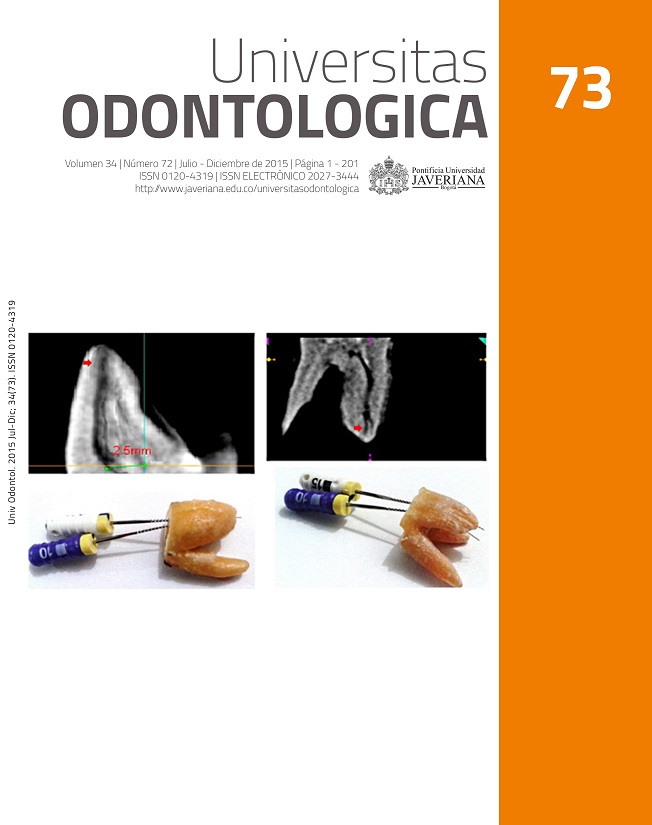
RESUMEN. Antecedentes: Las tasas de fracaso en los tratamientos de endodoncia se presentan en un mayor porcentaje en el primer molar superior debido a la no localización del conducto MV2 en la raíz MV. Los estudios sobre la morfología del primer molar superior no se pueden extrapolar a otras poblaciones debido a posibles diferencias étnicas. Objetivo: Determinar la incidencia de la configuración apical, la distancia media interorificio y la posible relación entre la distancia y la configuración apical de la raíz MV del primer molar superior en un grupo de población colombiana. Métodos: En este estudio descriptivo se analizó un total de 113 imágenes tomográficas computarizadas de haz de cono de la zona de molares superiores. El análisis consistió en la medición de la distancia entre los orificios de la raíz MV del primer molar superior a la altura del piso de la cámara pulpar para determinar si hay correlación con el tipo de configuración apical de la raíz MV. Resultados: La incidencia de configuraciones apicales II y IV fue del 41,59 % y 58,40 %, respectivamente. La distancia media entre los orificios de entrada de la raíz MV del primer molar superior para configuraciones apicales II y IV (clasificación Vertucci) fue 2,44 mm y 2,52 mm, respectivamente. Conclusiones: La configuración anatómica más común fue la tipo IV de Vertucci en 58,40 % de los casos. La distancia media interorificio en el grupo estudiado fue de 2,49 mm. No se encontró una relación entre la distancia interorificio y el tipo de configuración apical (p>0,05).
ABSTRACT. Background: Failure rates of endodontic treatments in maxillary first molar are mainly caused by the non-localization of the second mesiobuccal canal in the mesiobuccal root. Studies about upper first molar anatomy cannot be extrapolated from one population to another possibly because of ethnic variations. Objective: To determine the frequency of apical configuration, inter-orifice average distance, and possible relationship between the inter-orifice distance and the apical configuration of the maxillary first molar mesiobuccal root in a group of Colombians. Methods: In this descriptive study, 113 cone beam computed tomographic images of maxillary molar zones were analyzed. The study consisted of measuring, at the pulp chamber floor, the inter-orifice distance in maxillary first molars and determining apical configuration types of mesiobuccal roots. Measures included apical configuration, average inter-orifice distance, and possible association between the inter-orifice distance and the apical configuration (p=0.05). Data analysis was performed through CS software for 3D images. Results: The incidence of apical configurations types II and IV (Vertucci classification) was 41.59 % and 58.40 %, respectively. The average inter-orifice distances for apical configurations II and IV were 2.44 mm and 2.52 mm, respectively. Conclusions: The average inter-orifice distance was 2.49 mm. The most common anatomical configuration was IV in close to three fifths of the cases. There is no relationship between inter-orifice distance and apical configuration type.
Degerness RA, Bowles WR. Dimension, anatomy and morphology of the mesiobuccal root canal system in maxillary molars. J Endod. 2010; 36(6): 985-89.
Kabak Y, Abbott P. Prevalence of apical periodontitis and the quality of endodontic treatment in an adult Belarusian population. Int Endod J. 2005; 38(4): 238-45.
Weine FS, Healey HJ, Gerstein H, Evanson L. Canal configuration in the mesiobuccal root of the maxillary first molar and its endodontic significance. Oral Surg Oral Med Oral Pathol Oral Radiol Endod. 1969; 28(3): 419-25.
Park J-W, Lee J-K, Ha B-H, Choi J-H, Perinpanayagam H. Three-dimensional analysis of maxillary first molar mesiobuccal root canal configuration and curvature using micro–computed tomography. Oral Surg Oral Med Oral Pathol Oral Radiol Endod. 2009; 108(3): 437-42.
Smadi L, Khraisat A. Detection of a second mesiobuccal canal in the mesiobuccal roots of maxillary first molar teeth. Oral Surg Oral Med Oral Pathol Oral Radiol Endod. 2007; 103(3): e77-e81.
Witherspoon DE, Small JC, Regan JD. Missed canal systems are the most likely basis for endodontic retreatment of molars. Tex Dent J. 2013; 130(2): 127-39.
Blattner TC, George N, Lee CC, Kumar V, Yelton CD. Efficacy of cone-beam computed tomography as a modality to accurately identify the presence of second mesiobuccal canals in maxillary first and second molars: a pilot study. J Endod. 2010; 36(5): 867-70.
Baratto Filho F, Zaitter S, Haragushiku GA, de Campos EA, Abuabara A, Correr GM. Analysis of the internal anatomy of maxillary first molars by using different methods. J Endod. 2009; 35(3): 337-42.
Kim Y, Lee SJ, Woo J. Morphology of maxillary first and second molars analyzed by cone-beam computed tomography in a korean population: variations in the number of roots and canals and the incidence of fusion. J Endod. 2012; 38(8): 1063-8.
Campos Netto PA, Lins CCSA, Lins CV, Lima GA, Frazão MAG. Study of the internal morphology of the mesiobuccal root of upper first permanent molar using cone beam computed tomography. Int J Morphol. 2011; 29(2): 617-21.
Zheng Q-h, Wang Y, Zhou X-d, Wang Q, Zheng G-n, Huang D-m. A cone-beam computed tomography study of maxillary first permanent molar root and canal morphology in a Chinese population. J Endod. 2010; 36(9): 1480-4.
Vizzotto MB, Silveira PF, Arus NA, Montagner F, Gomes BP, da Silveira HE. CBCT for the assessment of second mesiobuccal (MB2) canals in maxillary molar teeth: effect of voxel size and presence of root filling. Int Endod J. 2013; 46(9): 870-6.
Domark JD, Hatton JF, Benison RP, Hildebolt CF. An ex vivo comparison of digital radiography and cone-beam and micro computed tomography in the detection of the number of canals in the mesiobuccal roots of maxillary molars. J Endod. 2013; 39(7): 901-5.
Bauman R, Scarfe W, Clark S, Morelli J, Scheetz J, Farman A. Ex vivo detection of mesiobuccal canals in maxillary molars using CBCT at four different isotropic voxel dimensions. Int Endod J. 2011; 44(8): 752-8.
Chang SW, Lee JK, Lee Y, Kum KY. In-depth morphological study of mesiobuccal root canal systems in maxillary first molars: review. Restor Dent Endod. 2013; 38(1): 2-10.
Cleghorn BM, Christie WH, Dong C. Root and root canal morphology of the human permanent maxillary first molar: a literature review. J Endod. 2006; 32(9): 813-21.
Spagnuolo G, Ametrano G, D’Antò V, Formisano A, Simeone M, Riccitiello F, Amato M, Rengo S. Microcomputed tomography analysis of mesiobuccal orifices and major apical foramen in first maxillary molars. Open Dent J. 2012; 6: 118-25.
Gu Y, Lee JK, Spångberg LSW, Lee Y, Park CM, Seo DG, Chang SW, Hur MS, Hong ST, Kum KY. Minimum-intensity projection for in-depth morphology study of mesiobuccal root. Oral Surg Oral Med Oral Pathol Oral Radiol Endod. 2011; 112(5): 671-7.
Zhang R, Yang H, Yu X, Wang H, Hu T, Dummer PMH. Use of CBCT to identify the morphology of maxillary permanent molar teeth in a Chinese subpopulation. Int Endod J. 2011; 44(2): 162-9.
Yamada M, Ide Y, Matsunaga S, Kato H, Nakagawa K. Three-dimensional analysis of mesiobuccal root canal of Japanese maxillary first molar using Micro-CT. Bull Tokyo Dent Coll. 2011; 52(2): 77-84.
Tuncer AK, Haznedaroglu F, Sert S. The location and accessibility of the second mesiobuccal canal in maxillary first molar. Europ J Dent. 2010; 4(1): 12.
Gilles J, Reader A. An SEM investigation of the mesiolingual canal in human maxillary first and second molars. Oral Surg Oral Med Oral Pathol Oral Radiol Endod. 1990; 70(5): 638-43.
Smadi L, Khraisat A. Root canal morphology of the mesiobuccal root in maxillary first molars of a Jordanian population. Gen Dent. 2006; 54(6): 413-6.
Ömer Görduysus M, Görduysus M, Friedman S. Operating microscope improves negotiation of second mesiobuccal canals in maxillary molars. J Endod. 2001; 27(11): 683-6.
Karaman GT, Onay EO, Ungor M, Colak M. Evaluating the potential key factors in assessing the morphology of mesiobuccal canal in maxillary first and second molars. Aust Endod J. 2011; 37(3): 134-40.
Weng X-L, Yu S-B, Zhao S-L, Wang H-G, Mu T, Tang R-Y, Zhou XD. Root canal morphology of permanent maxillary teeth in the Han nationality in Chinese Guanzhong area: a new modified root canal staining technique. J Endod. 2009; 35(5): 651-6.
Neelakantan P, Subbarao C, Subbarao CV. Comparative Evaluation of modified canal staining and clearing technique, cone-beam computed tomography, peripheral quantitative computed tomography, spiral computed tomography, and plain and contrast medium–enhanced digital radiography in studying root canal morphology. J Endod. 2010; 36(9): 1547-51.
Pattanshetti N, Gaidhane M, Al Kandari A. Root and canal morphology of the mesiobuccal and distal roots of permanent first molars in a Kuwait population–a clinical study. Int Endod J. 2008; 41(9): 755-62.
Kulild JC, Peters DD. Incidence and configuration of canal systems in the mesiobuccal root of maxillary first and second molars. J Endod. 1990; 16(7): 311-7.
Silva EJ, Nejaim Y, Silva AV, Haiter-Neto F, Cohenca N. Evaluation of root canal configuration of mandibular molars in a Brazilian population by using cone-beam computed tomography: an in vivo study. J Endod. 2013; 39(7): 849-52.
Reis AGdAR, Grazziotin-Soares R, Barletta FB, Fontanella VRC, Mahl CRW. Second canal in mesiobuccal root of maxillary molars is correlated with root third and patient age: a cone-beam computed tomographic study. J Endod. 2013; 39(5): 588-92.
Este periódico científico está registrado sob a licença Creative Commons Atribuição 4.0 Internacional. Portanto, este trabalho pode ser reproduzido, distribuído e comunicado publicamente em formato digital, desde que os autores e a Pontifícia Universidade Javeriana sejam reconhecidos. Citar, adaptar, transformar, autoarquivar, republicar e criar novas obras a partir do material é permitido para qualquer finalidade (mesmo comercial), desde que a autoria seja devidamente reconhecida, um link para o trabalho original seja fornecido e quaisquer alterações sejam indicadas. A Pontifícia Universidade Javeriana não detém os direitos sobre os trabalhos publicados, e o conteúdo é de exclusiva responsabilidade dos autores, que mantêm seus direitos morais, intelectuais, de privacidade e de publicidade.