
Cómo citar

Resumen
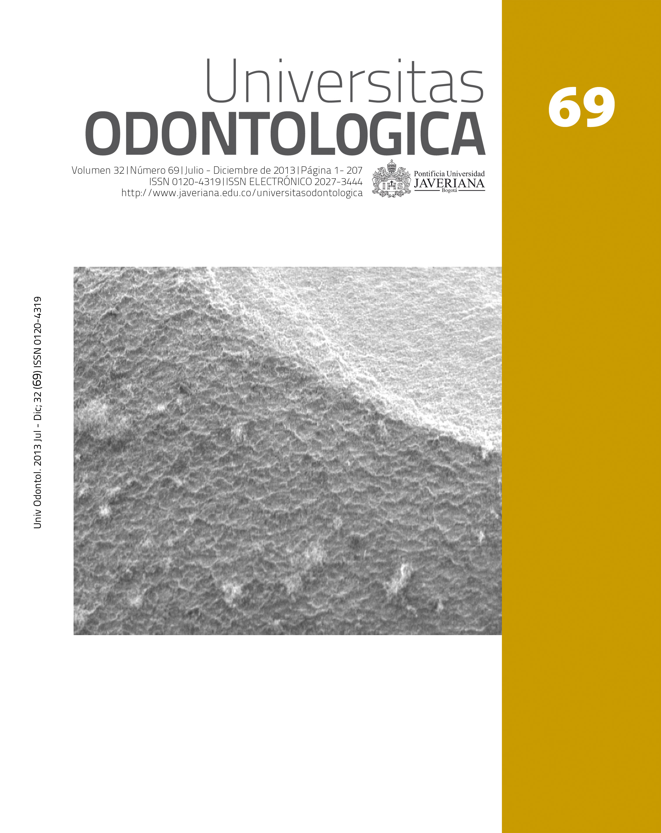
Antecedentes: Durante el proceso de morfogénesis dental se establecen complejas interfases morfofuncionales de unión entre los tejidos; así, entre el esmalte y la dentina se constituye la unión amelodentinaria (UAD), la cual corresponde a una interfase en la que elementos del esmalte y la dentina se interdigitan para conformar una solución de continuidad altamente resistente a las fuerzas verticales como una necesidad de los dientes para soportar el estrés funcional. Propósito: Describir la UAD a partir de los diferentes métodos de separación de la interfase entre el esmalte y la dentina y las técnicas de observación microscópica de dichas interfases reportadas en la literatura especializada. Métodos: Se realizó una revisión sistémica de la literatura mediante la búsqueda en las bases bibliográficas PubMed, Science-Direct, Hinari y SciELO con los descriptores enamel-dentin junction y enamel-dentin interface para clasificar los diferentes métodos de separación del esmalte y la dentina y la posterior observación y descripción de la UAD mediante diferentes técnicas de microscopia. Resultados: Los diferentes reportes evidencian que principalmente se han empleado técnicas que disuelven el esmalte con la acción de agentes ácidos, por lo que la descripción se ha centrado en la superficie de la UAD correspondiente a la dentina. Conclusiones: El patrón festoneado característico del espesor funcional de la UAD se puede observar de igual forma en dientes tratados con procedimientos químicos o separados con procedimientos físicos; emplear una de las dos obedece específicamente a la selección de la técnica de microscopia adecuada.
Background: Dental morphogenesis is an important phenomenon in which interaction between several tissues occurs. For instance, enamel and dentin form the dentine-enamel junction (DEJ), an interface that is highly resistant to vertical forces as the teeth is exposed to functional stress. Purpose: Describe the DEJ through several methods to separate the enamel and dentin interface and the techniques to observe microscopically such interfaces. Methods: We conducted a systematic review of the literature by searching the PubMed, Science-Direct, SciELO, and Hinari bibliographic databases with descriptors like enameldentin and enamel-dentin junction interface, in order to classify the different methods of separation of enamel and dentin and subsequent observation and description of the DEJ using different microscopy techniques. Results: Different physical and chemical techniques have been used to study the DEJ. The most frequent is enamel dissolution on acid, so that the description has focused on the surface of the DEJ corresponding to dentin. Conclusions: A scalloping pattern characteristic functional DEJ thickness can be observed similarly in teeth treated with chemical or physical processes and depending also the microscopy technique used.
Thesleff I, Nieminent P. Tooth morphogenesis and cell differentiation. Curr Op Cell Biol. 1996; 8: 844-50.
Arechaga J. The tooth as a model in organogenesis. Int J Dent Biol. 1995; 39: 13-23.
Thesleff I. Epithelial-mesenchymal signalling regulating tooth morphogenesis. J Cell Sci. 2003; 116: 1647-8.
Jernvall J, Thesleff I. Reiterative signaling and patterning during mammalian tooth morphogenesis. Mechanisms Dev. 2000; 92: 19-29.
Ten Cate AR. Histología oral: desarrollo, estructura y función. 2a ed. Buenos Aires: Panamericana; 1986.
Gómez de Ferrais ME, Campos A. Histología y embriología bucodental. 2a ed. Buenos Aires: Panamericana; 2002.
Garant PR. Oral cells and tissues. Chicago: Quintessence Books; 2003.
Neubueser A, Peters H, Balling R, Martin GR. Antagonistic interactions between FGF and BMP signaling pathways: a mechanism for positioning the sites of tooth formation. Cell. 1997; 90: 247-55.
Tucker AS, Sharpe PT. Molecular genetics of tooth morphogenesis and patterning: the right shape in the right place. J Dent Res. 1999; 78: 826-34.
Ortiz MA, Mejía CA. Actividad de los genes tipo msx-1 durante el desarrollo craneofacial. Rev Estomatol. 2007; 15: 34-8.
Vaahtokari A, Åberg T, Jernvall J, Keränen S, Thesleff I. The enamelknot as a signaling center in the developing mouse tooth. Mechanisms Dev. 1996; 54: 39-43.
Simmer JP, Hu J. Dental enamel formation and its impact on clinical dentistry. J Dent Educ. 2001; 65: 896-905.
Couve E. Ultrastructural changes during the life cycle of human odontoblast. Arch Oral Biol. 1986; 31: 643-51.
Mom M, Whittaker DA. Ultrastructure of the crown and root odontoblast. Ann Dent Univ Malaya. 2003; 10: 14-21.
Sisca RF, Provenza DV. Initial dentine formation in human deciduous teeth. Calc Tiss Res. 1972; 9: 1-26.
Peters H, Balling R. Teeth: where and how to make them. TIG. 1999; 15(29): 59-65.
Goldberg M, Septier D, Lecolle S, Chardin H, Quintana MA, Acevedo AC, Gafni G, Dillouya D, Vermelin L, Thonemann B, Schmalz G, Bissila-Mapahou P, Carreau JP. Dental mineralization. Int J Dev Biol. 1995; 39: 93-110.
Fincham AG, Moradian-Oldak J, Simmer JP. The structural biology of the developing dental enamel matrix. J Struct Biol. 1999; 126: 270-99.
Ruch JV, Lesot H, Begue-Kin C. Odontoblast differentiation. Int J Dev Biol. 1995; 39: 51-68.
Bodier-Houllé P, Steuer P, Meyer JM, Bigeard L, Cuisinier FJG. High-resolution electron-microscopic study of the relationship between human enamel and dentin crystals at the dentinoenamel junction. Cell and Tissue Research 2000; 301: 389-95.
Whittaker DA. The enamel-dentine junction of human and Macaca irus teeth: a light and electron microscopic study. J Anat. 1978; 125(2): 323-35.
Gallagher RR, Demos SG, Balooch M, Marshall GW, Marshall SJ. Optical spectroscopy and imaging of the dentin-enamel junction in human third molars. J Biomed Mater Res. 2003; 64A: 372-7.
Stock SR, Vieira AEM, Delbem ACB, Cannon ML, Xiao X, De Carlo F. Synchrotron microcomputed Tomography of the mature bovine dentinoenamel junction. J Struct Biol. 2008; 161(2): 162-71.
Chan YL, Ngan AHW, King NM. Nano-scale structure and mechanical properties of the human dentine-enamel junction. J Mech Behav Biomed Mater. 2011; 4(5): 785-95.
Sela J, Sela M, Lustmann J, Ulmansky M. Dentinoenamel Junction Area of a Resorbing Permanent Incisor Studied by Means of Scanning Electron Microscopy. J Dent Res. 1975; 54: 110-3.
Marshall SJ, Balooch M, Habelitz S, Balooch G, Gallagher G, Marshall W. The dentin-enamel junction. A natural multilevel interface. J Eur Ceramic Soc. 2003; 23: 2897-2904.
Lin CP, Douglas WH, Erlandsen SL. Scanning electron microscopy of Type I collagen at the dentin-enamel junction of human teeth. J Histochem Cytochem 1993; 41: 381-8.
Habelitz S, Marshall SJ, Marshall GW, Balooch M. The functional width of the dentino-enamel junction determined by AFM-based nanoscratching. J Struct Biol. 2001; 135: 294-301.
Imbeni V, Kruzic JJ, Marshal GW, Marshal SJ, Ritchie RO. The dentin-enamel junction and the fracture of human teeth. Nature Mater. 2005; 229-32.
Smith TM, Olejniczak AJ, Reid DJ, Ferrell RJ, Hublin JJ. Modern human molar enamel thickness and enamel-dentine junction shape. Arch Oral Biol. 2006; 51: 974-95.
Radlanski RJ, Renz H. Insular dentin formation pattern in human odontogenesis in relation to the scalloped dentino-enamel junction. Ann Anat. 2007; 189: 243-50.
Oliveira CA, Bergqvist LP, Line SR. A comparative analysis of the structure of the dentinoenamel junction in mammals. J Oral Sci. 2001; 43(4): 277-81.
Gil-Chavarría I, García-García R, Reyes-Gasga J. Comportamiento estructural de la unión esmalte- dentina en dientes humanos: un modelo mecánico-funcional. Acta Microscópica. 2008; 17(1): 34-47.
Brauer DS, Marshall GW, Marshall SJ. Variations in human DEJ scallop size with tooth type. J Dent. 2010; 38(7): 597-601.
Kraus BS. Morphologic relationships between enamel and dentin surfaces of lower first molar teeth. J Dent Res. 1952; 31: 248-56.
Rasmussen ST. Fracture properties of human teeth in proximity to the dentinoenamel function. J Dent Res. 1984; 63(11): 1279-83.
Moreno F, Mejía C. Comportamiento in vitro de la interface esmalte-dentina en premolares humanos sometidos a altas temperaturas. Trabajo de grado para optar por título de Magíster en Ciencias Biomédicas. Cali, Colombia: Escuela de Ciencias Básicas, Universidad del Valle; 2012.
Esta revista científica se encuentra registrada bajo la licencia Creative Commons Reconocimiento 4.0 Internacional. Por lo tanto, esta obra se puede reproducir, distribuir y comunicar públicamente en formato digital, siempre que se reconozca el nombre de los autores y a la Pontificia Universidad Javeriana. Se permite citar, adaptar, transformar, autoarchivar, republicar y crear a partir del material, para cualquier finalidad (incluso comercial), siempre que se reconozca adecuadamente la autoría, se proporcione un enlace a la obra original y se indique si se han realizado cambios. La Pontificia Universidad Javeriana no retiene los derechos sobre las obras publicadas y los contenidos son responsabilidad exclusiva de los autores, quienes conservan sus derechos morales, intelectuales, de privacidad y publicidad.
El aval sobre la intervención de la obra (revisión, corrección de estilo, traducción, diagramación) y su posterior divulgación se otorga mediante una licencia de uso y no a través de una cesión de derechos, lo que representa que la revista y la Pontificia Universidad Javeriana se eximen de cualquier responsabilidad que se pueda derivar de una mala práctica ética por parte de los autores. En consecuencia de la protección brindada por la licencia de uso, la revista no se encuentra en la obligación de publicar retractaciones o modificar la información ya publicada, a no ser que la errata surja del proceso de gestión editorial. La publicación de contenidos en esta revista no representa regalías para los contribuyentes.