

Palabras clave
odontología
odontología regenerativa
concentrados plaquetarios
fibrina rica en plaquetas
microscopía electrónica de barrido
Cómo citar

Resumen
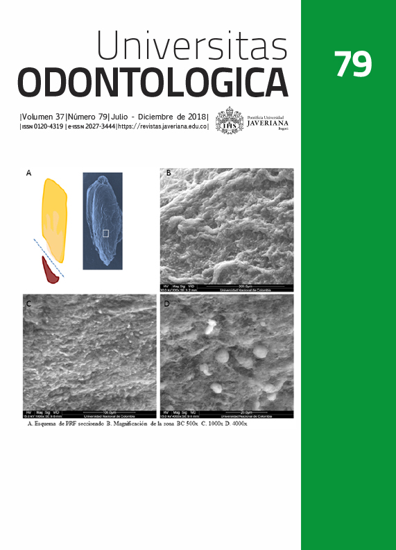
Antecedentes: La fibrina rica en plaquetas (PRF) es un concentrado plaquetario que se está usando con mayor frecuencia en medicina y odontología. Los resultados clínicos son variables posiblemente porque hay diferentes protocolos de obtención, equipos de centrifugado y técnicas de colocación. El desconocimiento de los aspectos estructurales puede afectar el resultado clínico. Objetivo: Describir las características estructurales de la PRF en las diferentes zonas de la membrana. Métodos: Se realizó un estudio experimental in vitro con 15 muestras de sangre periférica tomada de cinco voluntarios adultos, sanos, asistentes a la clínica odontológica de la Universidad Antonio, Popayán. Se hizo hemograma inicial, se recolectó sangre y se centrifugó (10 min x 3000 rpm). Las muestras se analizaron histológicamente y con microscopía electrónica de barrido (SEM). Se describió la estructura de la fibrina, las plaquetas y los leucocitos. Resultados: El promedio de recuento de plaquetas en sangre total fue de 251±31,74 x10³ x mm3 y en PRF fue de 832±123,43 x10³ x mm3. Macroscópicamente, se identificaron tres zonas del PRF: una superior con pocas plaquetas, una zona leucocitaria (BC) y una zona corpuscular roja. En el análisis de microscopía óptica muestra que en la zona BC hay mayor concentración plaquetaria. El análisis por SEM comprueba que la estructura de la red de fibrina y el contenido celular son diferenciales en cada zona. Conclusión: A partir del conocimiento estructural del PRF se pueden proponer aplicaciones que mejoren el rendimiento del material y por tanto los resultados clínicos.
Amrollahi P, Shah B, Seifi A, Tayebi L. Recent advancements in regenerative dentistry: A review. Mater Sci Eng C Mater Biol Appl. 2016; 69: 1383-90.
Choukroun J, Diss A, Simonpieri A, Girard MO, Schoeffler C, Dohan SL, et al. Platelet-rich fibrin (PRF): a second-generation platelet concentrate. Part IV: clinical effects on tissue healing. Oral Surg Oral Med Oral Pathol Oral Radiol Endod. 2006;101(3):e56-60.
Dohan Ehrenfest DM, Rasmusson L, Albrektsson T. Classification of platelet concentrates: from pure platelet-rich plasma (P-PRP) to leucocyte- and platelet-rich fibrin (L-PRF). Trends Biotechnol. 2009; 27(3): 158-67.
Borie E, Olivi DG, Orsi IA, Garlet K, Weber B, Beltran V, et al. Platelet-rich fibrin application in dentistry: a literature review. Int J Clin Exp Med. 2015; 8(5): 7922-9.
Tunali M, Ozdemir H, Kucukodaci Z, Akman S, Firatli E. In vivo evaluation of titanium-prepared platelet-rich fibrin (T-PRF): a new platelet concentrate. Br J Oral Maxillofac Surg. 2013; 51(5): 438-43.
Miron RJ, Fujioka-Kobayashi M, Bishara M, Zhang Y, Hernandez M, Choukroun J. Platelet-Rich Fibrin and Soft Tissue Wound Healing: A Systematic Review. Tissue Eng Part B Rev. 2017; 23(1): 83-99.
Dohan DM, Choukroun J, Diss A, Dohan SL, Dohan AJ, Mouhyi J, et al. Platelet-rich fibrin (PRF): a second-generation platelet concentrate. Part I: technological concepts and evolution. Oral Surg Oral Med Oral Pathol Oral Radiol Endod. 2006; 101(3): e37-44.
Dohan DM, Choukroun J, Diss A, Dohan SL, Dohan AJ, Mouhyi J, et al. Platelet-rich fibrin (PRF): a second-generation platelet concentrate. Part III: leucocyte activation: a new feature for platelet concentrates? Oral Surg Oral Med Oral Pathol Oral Radiol Endod. 2006; 101(3): e51-5.
Eren G, Gurkan A, Atmaca H, Donmez A, Atilla G. Effect of centrifugation time on growth factor and MMP release of an experimental platelet-rich fibrin-type product. Platelets. 2016; 27(5): 427-32.
Dohan Ehrenfest DM, Pinto NR, Pereda A, Jimenez P, Corso MD, Kang BS, et al. The impact of the centrifuge characteristics and centrifugation protocols on the cells, growth factors, and fibrin architecture of a leukocyte- and platelet-rich fibrin (L-PRF) clot and membrane. Platelets. 2017: 1-14.
Gutiérrez D, Restrepo A, Hinojosa J, Muñoz A, Ortiz Y. Centrifugation protocols to obtain platelet rich fibrin for dental applications. Innov Odontol. 2016; 1(1): 18-22.
Acosta P, Gutiérrez S, Bedoya M, García A, Moreno X. Evaluation of platelet-rich plasma effect at different times and concentrations on periodontal ligament fibroblast and osteoblast viability. Univ Odontol. 2017; 36(76): 1-23.
Dohan Ehrenfest DM. How to optimize the preparation of leukocyte- and platelet-rich fibrin (L-PRF, Choukroun's technique) clots and membranes: introducing the PRF Box. Oral Surg Oral Med Oral Pathol Oral Radiol Endod. 110. United States2010. p. 275-8; author reply 8-80.
Kumar YR, Mohanty S, Verma M, Kaur RR, Bhatia P, Kumar VR, et al. Platelet-rich fibrin: the benefits. Br J Oral Maxillofac Surg. 2015.
Verma UP, Yadav RK, Dixit M, Gupta A. Platelet-rich Fibrin: A Paradigm in Periodontal Therapy - A Systematic Review. J Int Soc Prev Community Dent. 2017; 7(5): 227-33.
Del Corso M, Vervelle A, Simonpieri A, Jimbo R, Inchingolo F, Sammartino G, et al. Current knowledge and perspectives for the use of platelet-rich plasma (PRP) and platelet-rich fibrin (PRF) in oral and maxillofacial surgery part 1: Periodontal and dentoalveolar surgery. Curr Pharm Biotechnol. 2012; 13(7): 1207-30.
Ranganathan AT, Chandran CR. Platelet-rich fibrin in the treatment of periodontal bone defects. J Contemp Dent Pract. 2014; 15(3): 372-5.
Thombre V, Koudale SB, Bhongade ML. Comparative evaluation of the effectiveness of coronally positioned flap with or without acellular dermal matrix allograft in the treatment of multiple marginal gingival recession defects. Int J Periodontics Restorative Dent. 2013; 33(3): e88-94.
Giraldo T, Sossa H. Endodoncia regenerativa: utilización de fibrina rica en plaquetas autóloga en dientes permanentes vitales con patología pulpar. Revisión narrativa de la literatura. Acta Odontológica colombiana [en línea]. 2014; 4(1): 91-112.
Faizuddin U, Solomon RV, Mattapathi J, Guniganti SS. Revitalization of traumatized immature tooth with platelet-rich fibrin. Contemp Clin Dent. 2015; 6(4): 574-6.
Dohan Ehrenfest DM, Del Corso M, Diss A, Mouhyi J, Charrier JB. Three-dimensional architecture and cell composition of a Choukroun's platelet-rich fibrin clot and membrane. J Periodontol. 2010; 81(4): 546-55.
Schar MO, Diaz-Romero J, Kohl S, Zumstein MA, Nesic D. Platelet-rich concentrates differentially release growth factors and induce cell migration in vitro. Clin Orthop Relat Res. 2015; 473(5): 1635-43.
Kobayashi E, Fluckiger L, Fujioka-Kobayashi M, Sawada K, Sculean A, Schaller B, et al. Comparative release of growth factors from PRP, PRF, and advanced-PRF. Clin Oral Investig. 2016; 20(9): 2353-2360.
Pinto G, Pigossi SC, Pessoa T, Nicoli LG, Araujo R, Marcantonio C, et al. Successful Use of Leukocyte Platelet-Rich Fibrin in the Healing of Sinus Membrane Perforation: A Case Report. Implant Dent. 2018. In press
Bhatsange A, Shende A, Deshmukh S, Japatti S. Management of fenestration using bone allograft in conjunction with platelet-rich fibrin. J Indian Soc Periodontol. 2017; 21(4): 337-40.
Johns DA, Shivashankar VY, Maroli RK, Joseph R. Invasive cervical root resorption: Engineering the lost tissue by regeneration. Contemp Clin Dent. 2013; 4(4): 536-9.
Kobayashi M, Kawase T, Horimizu M, Okuda K, Wolff LF, Yoshie H. A proposed protocol for the standardized preparation of PRF membranes for clinical use. Biologicals. 2012; 40(5): 323-9.
Moradian H, Rafiee A, Ayatollahi M. Design and Fabrication of a Novel Transplant Combined with Human Bone Marrow Mesenchymal Stem Cells and Platelet-rich Fibrin: New Horizons for Periodontal Tissue Regeneration after Dental Trauma. Iran J Pharm Res. 2017; 16(4): 1370-8.
Bilginaylar K. Uncommon Odontogenic Orocutaneous Fistula of the Jaw Treated with Platelet-Rich Fibrin. Case Rep Dent. 2017; 2017: 7174217.
Aoki N, Kanayama T, Maeda M, Horii K, Miyamoto H, Wada K, et al. Sinus Augmentation by Platelet-Rich Fibrin Alone: A Report of Two Cases with Histological Examinations. Case Rep Dent. 2016; 2016: 2654645.
Esta revista científica se encuentra registrada bajo la licencia Creative Commons Reconocimiento 4.0 Internacional. Por lo tanto, esta obra se puede reproducir, distribuir y comunicar públicamente en formato digital, siempre que se reconozca el nombre de los autores y a la Pontificia Universidad Javeriana. Se permite citar, adaptar, transformar, autoarchivar, republicar y crear a partir del material, para cualquier finalidad (incluso comercial), siempre que se reconozca adecuadamente la autoría, se proporcione un enlace a la obra original y se indique si se han realizado cambios. La Pontificia Universidad Javeriana no retiene los derechos sobre las obras publicadas y los contenidos son responsabilidad exclusiva de los autores, quienes conservan sus derechos morales, intelectuales, de privacidad y publicidad.
El aval sobre la intervención de la obra (revisión, corrección de estilo, traducción, diagramación) y su posterior divulgación se otorga mediante una licencia de uso y no a través de una cesión de derechos, lo que representa que la revista y la Pontificia Universidad Javeriana se eximen de cualquier responsabilidad que se pueda derivar de una mala práctica ética por parte de los autores. En consecuencia de la protección brindada por la licencia de uso, la revista no se encuentra en la obligación de publicar retractaciones o modificar la información ya publicada, a no ser que la errata surja del proceso de gestión editorial. La publicación de contenidos en esta revista no representa regalías para los contribuyentes.