Abstract
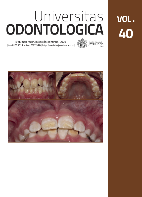
Background: Amelogenesis imperfecta (AI) is a hereditary condition that affects the structure of tooth enamel and causes sensitivity, predisposition to cavities, and psychological problems. In Colombia, its frequency, magnitude, distribution, and behavior are unknown, so it is necessary to carry out prevalence studies to implement preventive actions. Purpose: To determine the prevalence of AI in patients who have attended the Pontificia Universidad Javeriana clinics in Bogotá. Methods: A retrospective cross-sectional observational study was carried out, whose sample included 1,394 medical records of patients who attended between January 2015 and December 2017. Results: The prevalence of AI was 0.6 %, corresponding to 8 people affected, 4 men and 4 women between the ages of 9 and 10 years. The most frequent phenotype was hypoplastic in 7 patients (87.5 %) and one person had a hypocalcified phenotype (12.5 %). Taurodontism was the most frequent anomaly in the 8 patients (100 %). Seven of the eight patients (87.5 %) had a family history of AI. All the individuals had a lower-middle socioeconomic level and came from urban areas. Conclusions: This study is the first approximation to determine the prevalence of AI in a group of the Colombian population. Although the prevalence was low, it is comparable with the findings of other studies.
Wright J. The molecular etiologies and associated phenotypes of amelogenesis imperfecta. Am J Med Genet A. 2006 Dec 1; 140(23): 2547-2555.
Wright JT, Torain M, Long K, Seow K, Crawford P, Aldred MJ, Hart PS, Hart TC. Amelogenesis imperfecta: genotype-phenotype studies in 71 families. Cells Tissues Organs. 2011; 194(2-4): 279-83. https://doi.org/10.1159/000324339
Gadhia K, McDonald S, Arkutu N, Malik K. Amelogenesis imperfecta: an introduction. Br Dent J. 2012 Apr 27; 212(8): 377-379. https://doi.org/10.1038/sj.bdj.2012.314.
Wright JT, Hart PS, Aldred MJ, Seow K, Crawford PJ, Hong SP, Gibson CW, Hart TC. Relationship of phenotype and genotype in X-linked amelogenesis imperfecta. Connect Tissue Res. 2003; 44(Suppl 1): 72-78.
Hu JC, Yamakoshi Y. Enamelin and autosomal-dominant amelogenesis imperfecta. Crit Rev Oral Biol Med. 2003; 14(6): 387-398. https://doi.org/10.1177/154411130301400602
Rajpar MH, Harley K, Laing C, Davies RM, Dixon MJ. Mutation of the gene encoding the enamel-specific protein, enamelin, causes autosomal-dominant amelogenesis imperfecta. Hum Mol Genet. 2001 Aug 1; 10(16): 1673-1677. https://doi.org/10.1093/hmg/10.16.1673
Kim JW, Simmer JP, Hart TC, Hart PS, Ramaswami MD, Bartlett JD, Hu JC. MMP-20 mutation in autosomal recessive pigmented hypomaturation amelogenesis imperfecta. J Med Genet. 2005 Mar; 42(3): 271-5. https://doi.org/10.1136/jmg.2004.024505
Hart PS, Hart TC, Michalec MD, Ryu OH, Simmons D, Hong S, Wright JT. Mutation in kallikrein 4 causes autosomal recessive hypomaturation amelogenesis imperfecta. J Med Genet. 2004 Jul; 41(7): 545-549. https://doi.org/10.1136/jmg.2003.017657
Wang SK, Zhang H, Hu CY, Liu JF, Chadha S, Kim JW, Simmer JP, Hu JCC. FAM83H and autosomal dominant hypocalcified amelogenesis imperfecta. J Dent Res. 2021 Mar; 100(3): 293-301. https://doi.org/10.1177/0022034520962731.
Collins MA, Mauriello SM, Tyndall DA, Wright JT. Dental anomalies associated with amelogenesis imperfecta: a radiographic assessment. Oral Surg Oral Med Oral Pathol Oral Radiol Endod. 1999 Sep; 88(3): 358-364. https://doi.org/10.1016/s1079-2104(99)70043
Ravassipour DB, Powell CM, Phillips CL, Hart PS, Hart TC, Boyd C, Wright JT. Variation in dental and skeletal open bite malocclusion in humans with amelogenesis imperfecta. Arch Oral Biol. 2005 Jul; 50(7): 611-623. https://doi.org/10.1016/j.archoralbio.2004.12.003.
Seow WK. Taurodontism of the mandibular first permanent molar distinguishes between the tricho-dento-osseous (TDO) syndrome and amelogenesis imperfecta. Clin Genet. 1993 May; 43(5): 240-246. https://doi.org/10.1111/j.1399-0004.1993.tb03810.x
Zhang H, Koruyucu M, Seymen F, Kasimoglu Y, Kim JW, Tinawi S, Zhang C, Jacquemont ML, Vieira AR, Simmer JP, Hu JCC. WDR72 Mutations Associated with Amelogenesis Imperfecta and Acidosis. J Dent Res. 2019 May; 98(5): 541-548. https://doi.org/10.1177/0022034518824571
Witkop CJ Jr. Amelogenesis imperfecta, dentinogenesis imperfecta and dentin dysplasia revisited: problems in classification. J Oral Pathol. 1988 Nov; 17(9-10): 547-553. https://doi.org/10.1111/j.1600-0714.1988.tb01332.x
Sabandal MM, Schäfer E. Amelogenesis imperfecta: review of diagnostic findings and treatment concepts. Odontology. 2016 Sep; 104(3): 245-256. https://doi.org/10.1007/s10266-016-0266-1. Epub 2016 Aug 22.
Bäckman B. Amelogenesis imperfecta--clinical manifestations in 51 families in a northern Swedish county. Scand J Dent Res. 1988 Dec; 96(6): 505-516. https://doi.org/10.1111/j.1600-0722.1988.tb01590.x.
Chosack A, Eidelman E, Wisotski I, Cohen T. Amelogenesis imperfecta among Israeli Jews and the description of a new type of local hypoplastic autosomal recessive amelogenesis imperfecta. Oral Surg Oral Med Oral Pathol. 1979 Feb; 47(2): 148-156. https://doi.org/10.1016/0030-4220(79)90170-1.
Shokri A, Poorolajal J, Khajeh S, Faramarzi F, Kahnamoui HM. Prevalence of dental anomalies among 7- to 35-year-old people in Hamadan, Iran in 2012-2013 as observed using panoramic radiographs. Imaging Sci Dent. 2014 Mar; 44(1): 7-13. https://doi.org/10.5624/isd.2014.44.1.7.
Gutierrez SJ, Chaves M, Torres DM, Briceño I. Identification of a novel mutation in the enamalin gene in a family with autosomal-dominant amelogenesis imperfecta. Arch Oral Biol. 2007 May; 52(5): 503-506. https://doi.org/10.1016/j.archoralbio.2006.09.014
Calero J, Soto L. Amelogénesis imperfecta. Informe de tres casos en una familia en Cali, Colombia. Colomb Med. 2005 dec 1; 36(3): 47-50.
Simancas-Escorcia V, Berdal A, Diaz-Caballero A. Caracterización fenotípica del síndrome amelogénesis imperfecta–nefrocalcinosis: una revisión. Duazary 2019; 16(1): 129-143.
Gutiérrez S, Torres D, Briceño I, Gómez AM, Baquero E. Clinical and molecular analysis of the enamelin gene ENAM in Colombian families with autosomal dominant amelogenesis imperfecta. Genet Mol Biol. 2012 Jul; 35(3): 557-566. https://doi.org/10.1590/S1415-47572012000400003.
Gutiérrez SJ. Características clínicas de la caries en individuos con diferentes fenotipos de amelogénesis imperfecta. Univ Odontol. 2013 ene-jun; 32(68): 51-61
Duque Borrero AM, Rodríguez Manjarrez C, Soto Llanos OL, Triana Escobar FE. Prevalencia de anomalías dentales en pacientes de 4 a 14 años de edad, atendidos en las clínicas de odontopediatría de la Universidad del Valle en el período de enero de 2013 a junio de 2016. Gastrohnup. 2016 ene-abr 18; (1): 4-11.
Arias Acero NE, Fonseca Almánzar AM , Mora Oróstegui ML, Moreno Acuña DL, Pico Prada AR. Prevalencia de los defectos de desarrollo del esmalte en estudiantes de 7 a 9 años de dos instituciones educativas de Girón Santander 2014. Bucaramanga, Colombia: Repositorio Universidad Santo Tomas; 2015. https://repository.usta.edu.co/handle/11634/18755
Mafla AC, Córdoba DL, Rojas MN, Vallejos MA, Erazo MF, Rodríguez J. Prevalencia de defectos del esmalte dental en niños y adolescentes colombianos. Rev Fac Odontol Univ Antioq. 2014; 26(1): 106-125.
Rozo Gómez ER, Sánchez Castillo DZ, Silva Quevedo JD, Wong Arenas L. Prevalencia de hipoplasia e hipomineralización en niños de 7-13 años que asisten a la Clínica Odontopediátrica de la Universidad Cooperativa de Colombia. Bogotá, Colombia: Repositorio Universidad Cooperativa de Colombia; 2015. https://repository.ucc.edu.co/bitstream/20.500.12494/6489/1/2015-RozoySanchez-Prevalencia-Hipoplasia-Hipomineralizacion.pdf
Tovar S, Ziga E, Franco A, Jácome S, Ruiz III J. Estudio Nacional en Salud Bucal (ENSAB IV). Bogotá, Colombia: Ministerio de Salud; 2014.
Gupta SK, Saxena P, Jain S, Jain D. Prevalence and distribution of selected developmental dental anomalies in an Indian population. J Oral Sci. 2011 Jun; 53(2): 231-238. https://doi.org/10.2334/josnusd.53.231
Harini N, Don KR. Prevalence pattern of developmental anomalies of oral cavity in South Indian population-A hospital-based study. Drug Invention Today. 2019; 11(2).

This work is licensed under a Creative Commons Attribution 4.0 International License.
Copyright (c) 2021 Lizeth Paola Naranjo Jiménez, Myriam Adriana Muñoz Briceño, Ángela Suárez Castillo, Claudia Patricia Lamby Tovar, Sandra Janeth Gutierrez Prieto


