

Keywords
How to Cite

Abstract
Introduction: Congenital anomalies of the upper limb in the fetus or newborn include a wide spectrum of clinical presentations based on the penetrance and association between different anomalies. Therefore, their understanding and classification represents a real challenge for health professionals. The importance in understanding upper limb embryonic development events and the formation of its anatomical structures prevails on the fact it helps to fully understand the classification of congenital limb malformations. Generally, these malformations represent a potential functional loss in the newborn that when timely recognized can be submitted to surgical treatment, obtaining both aesthetic as well as functional satisfactory results. Methods: Fetal necropsies including autopsy data with their respective photographic documentation were performed from different Health centers in Bogotá, Colombia from 2012 to May 2019, which led to a review of the literature in this regard, clarifying the diagnostic criteria for congenital abnormalities of the upper limb including only review articles and case reports that facilitate their understanding, definition and classification. Results: Seven upper limb malformations are presented, with the elaboration of a manual to guide health professionals regarding diagnosis, timely recognition of congenital malformations of the upper limb in the fetus and newborn. The manual aims to strengthen malformation recognition for a timely interdisciplinary evaluation. Discussion: Congenital malformations of the upper limb comprise a challenge for health professionals, their understanding must be approached from the phenomena of embryonic limb development to understand, identify anatomical structures and diagnose in a timely manner.
2. Nielsen M, M.S., Esqueleto apendicular, in Atlas de Anatomía Humana2011, Panamericana Médica. p. 83.
3. Latarjet, R., Liard, Miembro superior, in Anatomía Humana2019, Panamericana Médica. p. 455-470.
4. Pró, E., in Anatomía Clínica2015, Panamericana Médica. p. 750 – 751.
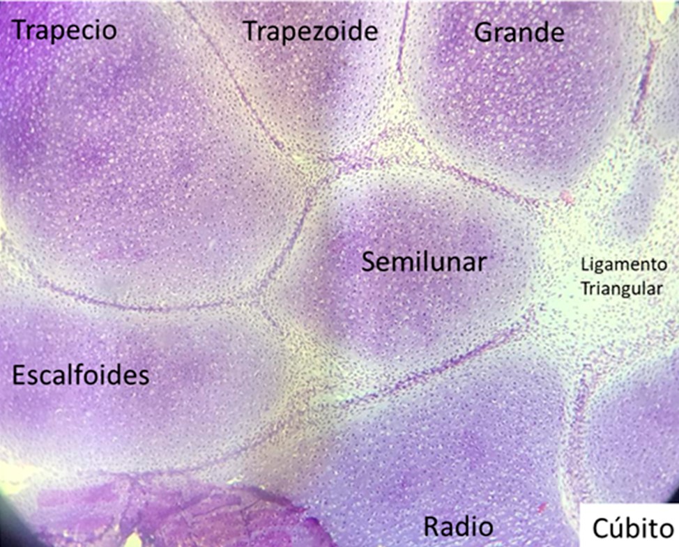
5. Moore, in Anatomía con orientación clínica2019, Wolters Kluwer. p. 771 – 779.
6. Carlson, B., Desarrollo de las extremidades, in Human embryology and Developmental Biology2014, Elsevier. p. 193- 215.
7. Lifemap, D. Lifemap Discovery Chapter 69. Development of the limbs 2020 [cited 2020 May 04, 2020]; Available from: https://discovery.lifemapsc.com/library/review-of-medical-embryology/chapter-69-development-of-the-limbs.
8. Al-Qattan, M.M., Y. Yang, and S.H. Kozin, Embryology of the upper limb. J Hand Surg Am, 2009. 34(7): p. 1340-50.
9. Tamura, K., et al., The autopod: its formation during limb development. Dev Growth Differ, 2008. 50 Suppl 1: p. S177-87.
10. Al-Qattan, M.M. and S.H. Kozin, Update on embryology of the upper limb. J Hand Surg Am, 2013. 38(9): p. 1835-44.
11. Cunningham, T.J. and G. Duester, Mechanisms of retinoic acid signalling and its roles in organ and limb development. Nat Rev Mol Cell Biol, 2015. 16(2): p. 110-23.
12. Gishen, K. and M. Askari, Congenital hand anomalies: etiology, classification, and treatment. J Craniofac Surg, 2014. 25(1): p. 284-94.
13. Dy, C.J., I. Swarup, and A. Daluiski, Embryology, diagnosis, and evaluation of congenital hand anomalies. Curr Rev Musculoskelet Med, 2014. 7(1): p. 60-7.
14. Zarante, I., et al., [Frequencies of congenital malformations: assessment and prognosis of 52,744 births in three cities of Colombia]. Biomedica, 2010. 30(1): p. 65-71.
15. Bermejo-Sanchez, E., et al., Phocomelia: a worldwide descriptive epidemiologic study in a large series of cases from the International Clearinghouse for Birth Defects Surveillance and Research, and overview of the literature. Am J Med Genet C Semin Med Genet, 2011. 157C(4): p. 305-20.
16. Swanson, A.B., A classification for congenital limb malformations. J Hand Surg Am, 1976. 1(1): p. 8-22.
17. Tonkin, M.A., et al., Classification of congenital anomalies of the hand and upper limb: development and assessment of a new system. J Hand Surg Am, 2013. 38(9): p. 1845-53.
18. Laub DR Jr, L.A., Hentz VR. , Plastic Surgery: Indications, Operations and Outcomes. , in Congenital hand anomalies2000. p. 1735Y1748.
19. Farr, S., et al., Peromelia - congenital transverse deficiency of the upper limb: a literature review and current prosthetic treatment. J Child Orthop, 2018. 12(6): p. 558-565.
20. Jain, S. and P.K. Lakhtakia, Profile of congenital transverse deficiencies among cases of congenital orthopaedic anomalies. J Orthop Surg (Hong Kong), 2002. 10(1): p. 45-52.
21. Mo., T., Pediatric orthopedics, in Congenital deformities1990, W.B. Saunders: Philadelphia, PA USA. p. 104–112.
22. Osadsky, C.R., Phocomelia: Case report and differential diagnosis. Radiol Case Rep, 2011. 6(4): p. 561.
23. de Jong, J.P., S.L. Moran, and S.K. Vilkki, Changing paradigms in the treatment of radial club hand: microvascular joint transfer for correction of radial deviation and preservation of long-term growth. Clin Orthop Surg, 2012. 4(1): p. 36-44.
24. Ekblom, A.G., T. Laurell, and M. Arner, Epidemiology of congenital upper limb anomalies in 562 children born in 1997 to 2007: a total population study from stockholm, sweden. J Hand Surg Am, 2010. 35(11): p. 1742-54.
25. Bayne, L.G. and M.S. Klug, Long-term review of the surgical treatment of radial deficiencies. J Hand Surg Am, 1987. 12(2): p. 169-79.
26. Colen, D.L., et al., Radial Longitudinal Deficiency: Recent Developments, Controversies, and an Evidence-Based Guide to Treatment. J Hand Surg Am, 2017. 42(7): p. 546-563.
27. Koskimies, E., et al., Congenital upper limb deficiencies and associated malformations in Finland: a population-based study. J Hand Surg Am, 2011. 36(6): p. 1058-65.
28. Manske, P.R. and C.A. Goldfarb, Congenital failure of formation of the upper limb. Hand Clin, 2009. 25(2): p. 157-70.
29. Sharma, A. and N. Sharma, A comprehensive functional classification of cleft hand: The DAST concept. Indian J Plast Surg, 2017. 50(3): p. 244-250.
30. Manske, P.R. and M.N. Halikis, Surgical classification of central deficiency according to the thumb web. J Hand Surg Am, 1995. 20(4): p. 687-97.
31. Watts AC, H.G. Congenital hand anomalies. in Mini Symposium: Children´s othopaedic surgery. 2006.
32. Miller, J.K., S.M. Wenner, and L.M. Kruger, Ulnar deficiency. J Hand Surg Am, 1986. 11(6): p. 822-9.
33. Bauer, A.S., M.S. Bednar, and M.A. James, Disruption of the radial/ulnar axis:congenital longitudinal deficiencies. J Hand Surg Am, 2013. 38(11): p. 2293-302; quiz 2302.
34. Malik, S., Syndactyly: phenotypes, genetics and current classification. Eur J Hum Genet, 2012. 20(8): p. 817-24.
35. Hay, S., Incidence of selected congenital malformations in Iowa. Am J Epidemiol, 1971. 94(6): p. 572-84.
36. Castilla, E.E., J.E. Paz, and I.M. Orioli-Parreiras, Syndactyly: frequency of specific types. Am J Med Genet, 1980. 5(4): p. 357-64.
37. Goodell, P.B., et al., Symbrachydactyly. Hand (N Y), 2016. 11(3): p. 262-270.
38. Baek, G.H. and H.J. Lee, Classification and surgical treatment of symphalangism in interphalangeal joints of the hand. Clin Orthop Surg, 2012. 4(1): p. 58-65.
39. Cushing, H., Hereditary Anchylosis of the Proximal Phalan-Geal Joints (Symphalangism). Genetics, 1916. 1(1): p. 90-106.
40. Singh, V., et al., Camptodactyly: An unsolved area of plastic surgery. Arch Plast Surg, 2018. 45(4): p. 363-366.
41. Choi, B.R., et al., Camptodactyly, arthropathy, coxa vara, pericarditis (CACP)syndrome: a case report. J Korean Med Sci, 2004. 19(6): p. 907-10.
42. McFarlane, R.M., et al., The anatomy and treatment of camptodactyly of the small finger. J Hand Surg Am, 1992. 17(1): p. 35-44.
43. Flatt, A.E., The troubles with pinkies. Proc (Bayl Univ Med Cent), 2005. 18(4): p. 341-4.
44. Leung, A.K. and C.P. Kao, Familial clinodactyly of the fifth finger. J Natl Med Assoc, 2003. 95(12): p. 1198-200.
45. Tsai, J., Congenital radioulnar synostosis. Radiol Case Rep, 2017. 12(3): p. 552-554.
46. Cleary, J.E. and G.E. Omer, Jr., Congenital proximal radio-ulnar synostosis. Natural history and functional assessment. J Bone Joint Surg Am, 1985. 67(4): p. 539-45.
47. Hansen, O.H. and N.O. Andersen, Congenital radio-ulnar synostosis. Report of 37 cases. Acta Orthop Scand, 1970. 41(3): p. 225-30.
48. Ahmed, H., et al., Genetic Overview of Syndactyly and Polydactyly. Plast Reconstr Surg Glob Open, 2017. 5(11): p. e1549.
49. Abzug, J.M. and S.H. Kozin, Treatment of postaxial polydactyly type B. J Hand Surg Am, 2013. 38(6): p. 1223-5.
50. Malik, S., Polydactyly: phenotypes, genetics and classification. Clin Genet, 2014. 85(3): p. 203-12.
51. Tomaszewski, R. and A. Bulandra, Ulnar dimelia-diagnosis and management of a rare congenital anomaly of the upper limb. J Orthop, 2015. 12(Suppl 1): p. S121-4.
52. Chinegwundoh, J.O., M. Gupta, and W.A. Scott, Ulnar dimelia. Is it a true duplication of the ulna? J Hand Surg Br, 1997. 22(1): p. 77-9.
53. Jameel, J., et al., Ulnar dimelia variant: a case report. J Orthop Traumatol, 2011. 12(3): p. 163-5.
54. Al-Qattan, M.M., et al., Classification of the mirror hand-multiple hand spectrum. J Hand Surg Br, 1998. 23(4): p. 534-6.
55. Gaba, S., et al., Mirror Hand: An Uncommon Neglected Case Managed with Pollicisation. World J Plast Surg, 2017. 6(2): p. 263-265.
56. Dell, P.C., Macrodactyly. Hand Clin, 1985. 1(3): p. 511-24.
57. Barsky, A.J., Macrodactyly. J Bone Joint Surg Am, 1967. 49(7): p. 1255-66.
58. Bisneto, E.N., Congenital Deformities of the Upper Limbs.: Part I: Failure of Formation. Rev Bras Ortop, 2012. 47(5): p. 545-52.
59. Bisneto, E.N.F., Congenital Deformities of the Upper Limbs. Part III: Overgrowth; Undergrowth; Streeter and Others. Rev Bras Ortop, 2013. 48(2): p. 121-125.
60. Miura, T., S. Torii, and R. Nakamura, Brachymetacarpia and brachyphalangia. J Hand Surg Am, 1986. 11(6): p. 829-36.
61. Sayadi, L., M. Chopan, and D. Laub, Thumb Hypoplasia. Eplasty, 2015. 15: p. ic62.
62. James, M.A., H.R. McCarroll, Jr., and P.R. Manske, Characteristics of patients with hypoplastic thumbs. J Hand Surg Am, 1996. 21(1): p. 104-13.
63. Blauth, W., [The hypoplastic thumb]. Arch Orthop Unfallchir, 1967. 62(3): p. 225-46.
64. Tonkin, M., Surgical reconstruction of congenital thumb hypoplasia. Indian J Plast Surg, 2011. 44(2): p. 253-65.
65. Walter, J.H., Jr., L.R. Goss, and A.T. Lazzara, Amniotic band syndrome. J Foot Ankle Surg, 1998. 37(4): p. 325-33.
66. Paladini, D., et al., Congenital constriction band of the upper arm: the role of three-dimensional ultrasound in diagnosis, counseling and multidisciplinary consultation. Ultrasound Obstet Gynecol, 2004. 23(5): p. 520-2.
67. Shetty, P., et al., Amniotic band syndrome. Indian J Surg, 2013. 75(5): p. 401-2.

This work is licensed under a Creative Commons Attribution 4.0 International License.
Copyright (c) 2020 Maria Lucia Gutierrez, Diana Carolina Sarmiento Osorio, Javier Fabricio Guillén Olaya, Jorge Andrés Franco Zuluaga